. 2013 Dec;41(22):10298-311.
doi: 10.1093/nar/gkt802. Epub 2013 Sep 5.

Co-operation of BRCA1 and POH1 relieves the barriers posed by 53BP1 and RAP80 to resection

Affiliations

Co-operation of BRCA1 and POH1 relieves the barriers posed by 53BP1 and RAP80 to resection

Andreas Kakarougkas et al. Nucleic Acids Res. 2013 Dec.

Abstract

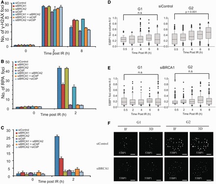
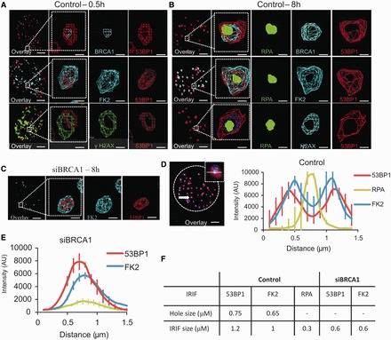
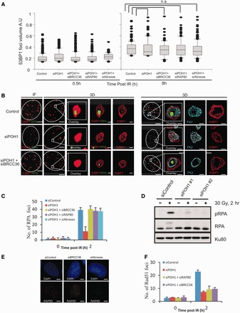
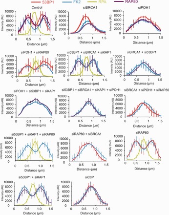
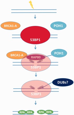
In G2 phase cells, DNA double-strand break repair switches from DNA non-homologous end-joining to homologous recombination. This switch demands the promotion of resection. We examine the changes in 53BP1 and RAP80 ionizing radiation induced foci (IRIF) in G2 phase, as these are factors that restrict resection. We observed a 2-fold increase in the volume of 53BP1 foci by 8 h, which is not seen in G1 cells. Additionally, an IRIF core devoid of 53BP1 arises where RPA foci form, with BRCA1 IRIF forming between 53BP1 and replication protein A (RPA). Ubiquitin chains assessed using α-FK2 antibodies are similarly repositioned. Repositioning of all these components requires BRCA1's BRCT but not the ring finger domain. 53BP1, RAP80 and ubiquitin chains are enlarged following POH1 depletion by small interfering RNA, but a devoid core does not form and RPA foci formation is impaired. Co-depletion of POH1 and RAP80, BRCC36 or ABRAXAS allows establishment of the 53BP1 and ubiquitin chain-devoid core. Thus, the barriers posed by 53BP1 and RAP80 are relieved by BRCA1 and POH1, respectively. Analysis of combined depletions shows that these represent distinct but interfacing barriers to promote loss of ubiquitin chains in the IRIF core, which is required for subsequent resection. We propose a model whereby BRCA1 impacts on 53BP1 to allow access of POH1 to RAP80. POH1-dependent removal of RAP80 within the IRIF core enables degradation of ubiquitin chains, which promotes loss of 53BP1. Thus, POH1 represents a novel component regulating the switch from non-homologous end-joining to homologous recombination.


Figures

Figure 1.
Figure 1.
BRCA1 functions downstream of CtIP in G2 to promote resection and 53BP1 repositioning. (A) A549 cells were exposed to 3 Gy IR and γH2AX foci enumerated to 8 h post-IR. G2 cells were identified by CENPF staining (4). RPA (B) andRAD51 (C) foci in G2 phase A549 cells post 3 Gy IR following treatment with the indicated siRNAs. (D and E) Analysis of 53BP1 foci volume in A549 cells following 3 Gy IR in untreated cells (D) and following treatment with siRNA BRCA1 (E). (F) Representative images of nuclei immunostained for 53BP1 at 8 h after 3 Gy IR in G1 and G2 with or without siRNA BRCA1. Scale bars—5 μm. BRCA1 knockdown efficiency is shown in Supplementary Figure S1.
Figure 2.
Figure 2.
BRCA1 promotes 53BP1 repositioning in G2 creating a core devoid of 53BP1 and ubiquitin chains. (A and B) Analysis of G2 A549 cells at 0.5 (A) and 8 (B) h post 3 Gy IR. Following immunostaining with the indicated antibodies, 3D IRIF analysis was undertaken. The red and green signals represent 53BP1 and RPA, respectively; the blue signal is as indicated. Here, and in all cases where four antibodies were used, DAPI was omitted. This allowed us to use a blue secondary antibody (Alexa 350) for identifying G2 phase cells. A typical example would be as follows: Rabbit CENPF antibody coupled with a blue secondary antibody, rat RPA antibody coupled with a green secondary antibody, Rabbit BRCA1 antibody (these foci are also visible in the blue channel but do not interfere with pan-nuclear appearance of CENPF) coupled with a red secondary antibody, mouse 53BP1 coupled with a far red secondary antibody. ‘Wireframe’ images are displayed allowing 3D visualization. Foci volume enlarges from 0.5 to 8 h post-IR generating an expanded core lacking 53BP1. BRCA1 localizes internally to 53BP1 and RPA lies within the core. (C) Images following siRNA BRCA1 8 h post-IR. Scale bars—5 and 0.5 μm in magnified images. (D and E) Quantification of fluorescence intensity profiles along a line drawn through the centre of IRIF at 8 h post-irradiation. In control cells (D) by 8 h post-IR, 53BP1 and FK2 IRIF are distributed in a bipolar manner whilst in BRCA1 k.d cells, single peaks persist. A minimum of 10 cells were analysed from each of three independent experiments. Error bars represent SD. The 53BP1 and RPA foci intensity assessed in (E) represents an unbiased average of all IRIF after siRNA BRCA1 (i.e. those with or without detectable RPA foci). Analysis of IRIF with RPA foci is shown in Supplementary Figure S3. The 53BP1 repositioning was impaired to the same extent in IRIF with or without RPA foci after siRNA BRCA1. (F) Quantification of IRIF clearance and overall size in Control and BRCA1 siRNA-treated cells. IRIF clearance represents the distance between the peaks in IRIF that show a bipolar distribution, whereas the foci size was determined by measuring the distance between the outer edges of the peaks at 50% intensity. 53BP1 knockdown efficiency is shown in Supplementary Figure S1. The size of the RPA peak was not estimated after siRNA BRCA1 due to the lack of a defined peak.
Figure 3.
Figure 3.
The BRCT but not the RING domain of BRCA1 is required for resection, RAD51 loading and 53BP1 repositioning during HR in G2. (A) Brca1FH-WT/FH-WT, Brca1FH-I26A/FH-I26A and Brca1S1598F/S1598F MEFs were irradiated with 3 Gy IR in the presence of aphidocolin. Cells were harvested 8 h post-IR and immunostained with DAPI, RPA and p-H3 antibodies to identify G2 cells. (B) As for (A), but RAD51 foci were enumerated 2 h post 3 Gy IR. (C) Analysis of 53BP1 foci volume in G2 phase Brca1FH-WT/FH-WT, Brca1FH-I26A and Brca1FH-S1598F MEFs 0.5 and 8 h following 3Gy IR. Analysis was carried out as in Figure 1C. (D) Quantification of the intensity of 53BP1 in G2 phase Brca1FH-WT/FH-WT, Brca1FH-I26A and Brca1FH-S1598F MEFs. Analysis was carried out as in Figure 2D. A minimum of 10 cells were analysed from three independent experiments. Error bars represent SD.
Figure 4.
Figure 4.
POH1 promotes resection in G2 by overcoming the inhibitory barriers of 53BP1 and RAP80. (A) 53BP1 foci volume was estimated in A549 cells treated with the indicated siRNA as in Figure 1C. (B) A549 cells were immunostained with the indicated antibodies 8 h post 3 Gy. The far left panels are a projection of the immunofluorescence (IF) Z-stacked images. The 3D model conversions of these images are shown in the subsequent panels. A devoid core of 53BP1 or FK2 does not form following siRNA POH1 but is visible following siRNA BRCC36 + POH1. A single oligonucleotide to POH1 was used, but similar results were obtained using a pool of siRNA POH1 oligonucleotides. RPA (C) and Rad51 (F) foci were enumerated in A549 cells treated with the indicated siRNA, and exposed to 3 Gy. (D) Resection was also assessed by monitoring phosphorylation of RPA on Ser 4 and 8 by western blotting. A549 cells were irradiated with 30 Gy following siRNA transfection, and whole-cell extracts were prepared at 2 h post-IR. (E) Depletion of BRCC36 or Abraxas results in loss of RAP80 demonstrating that they are required for RAP80 stability. Knockdown efficiency following siRNA POH1 is shown in Supplementary Figure S1. The 53BP1 foci size was assessed in all G2 cells following siRNA POH1, regardless of whether they have RPA foci or not. Scale bars—5 μm and 0.5 μm in magnified images.
Figure 5.
Figure 5.
Analysis of IRIF clearance from resection sites in G2 phase following combined siRNA treatments. Clearance of 53BP1, RAP80 and Ubiquitin chains from the IRIF core is required for resection to proceed normally. Here, the distribution of these factors is demonstrated by quantification of the fluorescence intensity profiles along a line drawn through the centre of IRIF at 8 h post-irradiation (as in F). RPA foci intensity was not assessed in those situations where foci numbers were substantially (<50%) decreased due inaccuracy in assessing the average intensity of two distinct populations. The requirement of various factors for the clearance of 53BP1, RAP80 and Ubiquitin chains was analysed using combined siRNA-mediated knockdown. Knockdown efficiency following multiple siRNA transfection is shown in Supplementary Figure S1. A minimum of 10 cells were analysed from three independent experiments. Error bars represent SD. Quantification of IRIF clearance and overall size in cells treated with the various combinations of siRNA-mediated knockdown is shown in Supplementary Figure S6. The data for control cells and siRNA BRCA1 for 53BP1, ubiquitin chains and RPA foci is the same as that shown in Figure 2D and E. RAP80 analysis is additional.
Figure 6.
Figure 6.
Model showing the removal of the inhibitory barriers posed to resection by 53BP1, RAP80 and Ubiquitin chains. Following the induction of DSBs, several factors including 53BP1, BRCA1 and RAP80 rapidly accumulate and form IRIF. The recruitment of 53BP1 and BRCA1 is dependent on the formation of ubiquitin chains at DSBs, which also form IRIF and can be visualized by antibodies that detect conjugated ubiquitin (a-FK2). Three complexes (BRCA1A-C) involving BRCA1 have been described (16). BRCA1-A comprises ABRAXAS, RAP80, BRCC36, BRCC45 and MERIT40. The presence of 53BP1, RAP80 and ubiquitin chains promote repair via NHEJ, as they are inhibitory to HR by blocking the process of resection. In G2 phase, the inhibitory barrier posed by these factors needs to be overcome for resection to proceed. Initially, BRCA1, via a process that requires its BRCT domain, ‘primes’ 53BP1 to allow POH1 access to RAP80. In the absence of BRCA1, both 53BP1 and RAP80 remain in the IRIF core. POH1 promotes the clearance of RAP80, which in turn allows removal of the ubiquitin chains from the IRIF core possibly via a DUB activity. This, in turn, promotes the full clearance of 53BP1 from the IRIF core. Thus, BRCA1 alone is not sufficient to promote clearance of 53BP1 from the core; POH1 is additionally required. Likewise, BRCA1 and POH1 are required to promote the clearance of RAP80 from the core. Once all these factors have been cleared from the IRIF core, nucleases can promote 5′-3′ resection, thus allowing repair by HR to proceed. We have proposed a linear feedback model to accommodate the finding that depletion of BRCA1 or POH1 can block clearance of both RAP80 and 53BP1, demonstrating that they are inter-dependent barriers. In the model shown, we have only depicted the removal of proteins/modifications from the IRIF core and have not included the repositioning of the proteins to the periphery of the foci.

Similar articles

Cited by

References

    1. Mahaney BL, Meek K, Lees-Miller SP. Repair of ionizing radiation-induced DNA double-strand breaks by non-homologous end-joining. Biochem. J. 2009;417:639–650. - PMC - PubMed
    1. Petermann E, Helleday T. Pathways of mammalian replication fork restart. Nat. Rev. Mol. Cell Biol. 2010;11:683–687. - PubMed
    1. Beucher A, Birraux J, Tchouandong L, Barton O, Shibata A, Conrad S, Goodarzi AA, Krempler A, Jeggo PA, Lobrich M. ATM and Artemis promote homologous recombination of radiation-induced DNA double-strand breaks in G2. EMBO J. 2009;28:3413–3427. - PMC - PubMed
    1. Shibata A, Conrad S, Birraux J, Geuting V, Barton O, Ismail A, Kakarougkas A, Meek K, Taucher-Scholz G, Lobrich M, et al. Factors determining DNA double-strand break repair pathway choice in G2 phase. EMBO J. 2011;30:1079–1092. - PMC - PubMed
    1. Panier S, Durocher D. Regulatory ubiquitylation in response to DNA double-strand breaks. DNA Repair (Amst.) 2009;8:436–443. - PubMed

Publication types

MeSH terms