. 2013 Dec;41(22):10488-508.
doi: 10.1093/nar/gkt803. Epub 2013 Sep 5.

Suppression of HPV-16 late L1 5'-splice site SD3632 by binding of hnRNP D proteins and hnRNP A2/B1 to upstream AUAGUA RNA motifs

Affiliations

Suppression of HPV-16 late L1 5'-splice site SD3632 by binding of hnRNP D proteins and hnRNP A2/B1 to upstream AUAGUA RNA motifs

Xiaoze Li et al. Nucleic Acids Res. 2013 Dec.

Abstract

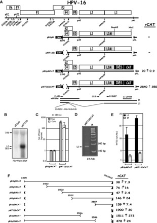
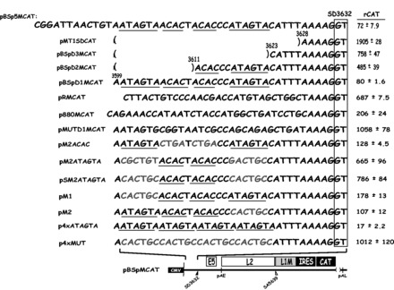
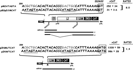
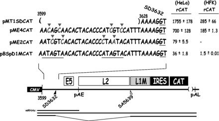
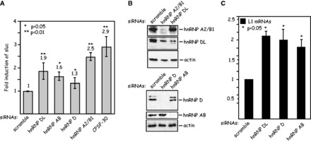
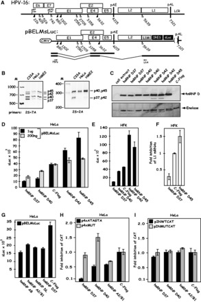
Human papillomavirus type 16 (HPV-16) 5'-splice site SD3632 is used exclusively to produce late L1 mRNAs. We identified a 34-nt splicing inhibitory element located immediately upstream of HPV-16 late 5'-splice site SD3632. Two AUAGUA motifs located in these 34 nt inhibited SD3632. Two nucleotide substitutions in each of the HPV-16 specific AUAGUA motifs alleviated splicing inhibition and induced late L1 mRNA production from episomal forms of the HPV-16 genome in primary human keratinocytes. The AUAGUA motifs bind specifically not only to the heterogeneous nuclear RNP (hnRNP) D family of RNA-binding proteins including hnRNP D/AUF, hnRNP DL and hnRNP AB but also to hnRNP A2/B1. Knock-down of these proteins induced HPV-16 late L1 mRNA expression, and overexpression of hnRNP A2/B1, hnRNP AB, hnRNP DL and the two hnRNP D isoforms hnRNP D37 and hnRNP D40 further suppressed L1 mRNA expression. This inhibition may allow HPV-16 to hide from the immune system and establish long-term persistent infections with enhanced risk at progressing to cancer. There is an inverse correlation between expression of hnRNP D proteins and hnRNP A2/B1 and HPV-16 L1 production in the cervical epithelium, as well as in cervical cancer, supporting the conclusion that hnRNP D proteins and A2/B1 inhibit HPV-16 L1 mRNA production.


Figures

Figure 1.
Figure 1.
(A). Identification of splicing inhibitory sequences immediately upstream of HPV-16 late 5′-splice SD3632. Schematic representation of the HPV-16 genome and the subgenomic HPV-16 expression plasmids. The early and late viral promoters p97 and p670 are indicated. Numbers indicate nucleotide positions of 5′- (filled triangles) and 3′-splice sites (open triangles). The early and late poly (A) sites named pAE and pAL are indicated. L1M represents a previously described mutant HPV-16 L1 sequence in which a number of nucleotide substitutions that inactivate splicing silencers have been inserted downstream of SA5639 (29,32). The sequence of the HPV-16 late 5′-splice site SD3632 is shown. IRES, the poliovirus IRES sequence; CAT, CAT reporter gene; CMV, human cytomegalovirus immediate early promoter; U, unspliced mRNA. Restriction sites BamHI and XhoI used for insertion of IRES and CAT are indicated. mRNAs produced by the plasmids are indicated. The position of the L1 northern blot probe and RT-PCR primers (arrows) are indicated. CAT protein levels produced by each CAT plasmid in the transfected HeLa cells are shown to the right. rCAT was calculated as described in ‘Materials and Methods’ section. Mean values and standard deviations are shown. (B) Northern blot on cytoplasmic RNA extracted from HeLa cells transfected with pBSpM or pMT1SD and probed with the L1 probe. (C) Real-time PCR of spliced HPV-16 L1 mRNA in nuclear (Nuc) or cytoplasmic (Cyto) fractions of transfected HeLa cells using primers Pr681 and Pr5687 mRNAs as described in ‘Materials and Methods’ section. Graph displays fold difference in L1 mRNA levels between pBSpMCAT and pMT1SDCAT. (D) RT-PCR with primers Pr681 and Pr5687 on cDNA of cytoplasmic RNA extracted from HeLa cells transfected with pMT1SDCAT. (E) Real-time PCR of HPV-16 spliced L1 mRNAs or unspliced (U) mRNA in nuclear (Nuc) or cytoplasmic (Cyto) fractions of transfected HeLa cells using primers Pr681, L2S and Pr5687 mRNAs as described in ‘Materials and Methods’ section. Graph displays the ratio between cytoplasmic and nuclear L1 or unspliced mRNAs produced from pBSpMCAT and pMT1SDCAT. (F) Schematic representations of deletions introduced in HPV-16 subgenomic expression plasmid pBSpMCAT. Plasmid names are shown to the left. Lines represent HPV-16 sequences present in the various plasmids, and numbers indicate ends of deletions. CAT protein levels produced by each plasmid in the transfected cells are shown to the right. rCAT was calculated as described in ‘Materials and Methods’ section. Mean values and standard deviations are shown.
Figure 2.
Figure 2.
Two nucleotide substitutions in each AUAGUA motif inactivate the splicing suppressors at HPV-16 late 5′-splice site SD3632. Schematic representations of subgenomic HPV-16 expression plasmids. Plasmid names are shown to the left. HPV-16 sequences present in the various plasmids are shown. Nucleotides in red indicate mutated positions. The AUAGUA and ACAC motifs are underlined. Filled and empty triangles represent late 5′-splice site SD3632 and late 3′-splice site SA5639. Early and late poly (A) signals pAE and pAL are indicated. L1M, a mutant L1 sequence in which splicing silencers downstream of SA5639 had been inactivated (29,32); CMV, human cytomegalovirus immediate early promoter; IRES, the poliovirus IRES sequence; CAT, CAT reporter gene. CAT protein levels produced by each plasmid in the transfected cells are shown to the right. rCAT was calculated as described in ‘Materials and Methods’ section. Mean values and standard deviations are shown.
Figure 3.
Figure 3.
The splicing silencer at HPV-16 5′-splice site SD3632 primarily inhibits production of spliced HPV-16 L1 mRNAs. The mutant or wild-type splicing silencer was inserted in a ‘pre-spliced’ L1 mRNA made from pBSpMCAT generating plasmids pINMUTCAT and pINWTCAT to monitor the effect of the silencer on an L1 mRNA that cannot be spliced. The effect was compared with the CAT protein levels produced from plasmids pM2ATAGTA and pBSpD1MCAT that contain the same wild-type and mutant splicing silencer sequences as the ‘pre-spliced’ mRNAs produced from produced pINMUTCAT and pINWTCAT. CAT levels produced by each plasmid in the transfected cells are shown to the right. rCAT was calculated as described in ‘Materials and Methods’ section. Mean values and standard deviations are shown. The ratio between the CAT levels produced from mutant and wild-type plasmids are shown to the right. CMV, human cytomegalovirus immediate early promoter; IRES, the poliovirus IRES sequence; CAT, CAT reporter gene. mRNAs produced by the HPV plasmids are indicated.
Figure 4.
Figure 4.
Two nucleotide substitutions in each AUAGUA motif inactivate the splicing suppressors at HPV-16 late 5′-splice site SD3632. Schematic representations of subgenomic HPV-16 expression plasmids. Plasmid names are shown to the left. HPV-16 sequences present in the various plasmids are shown. Nucleotides in red marked with red triangles indicate mutated positions. The AUAGUA motifs are underlined. Filled and empty triangles represent late 5′-splice site SD3632 and late 3′-splice site SA5639. Early and late poly (A) signals pAE and pAL are indicated. L1M, a mutant L1 sequence in which splicing silencers downstream of SA5639 had been inactivated (29,32); CMV, human cytomegalovirus immediate early promoter; IRES, the poliovirus IRES sequence; CAT, CAT reporter gene. mRNAs produced by the HPV-16 plasmids are indicated. CAT protein levels produced by each plasmid in transfected HeLa cells or HFKs cells are shown to the right. rCAT was calculated as described in ‘Materials and Methods’ section. Mean values and standard deviations are shown.
Figure 5.
Figure 5.
(A). The two AUAGUA motifs inhibit late L1 mRNA splicing in the full-length, episomal HPV-16 genome expressed in human primary keratinocytes. Structure of the HPV-16 genomic plasmids pHPV16ANsL. LoxP sites and HPV-16 early (p97) and late (p670) promoters are indicated. The cassette encoding the Rous sarcoma virus long terminal repeat promoter driving the neomycin resistance gene, followed by the simian virus 40 polyA signal is indicated (RSVneo). Arrows indicate positions of PCR primers 16S and 16A. The effect of the cre recombinase produced by the cotransfected plasmid pCAGSS-nlscre on these plasmids is illustrated. pAE and pAL, HPV-16 early and late polyA signals, respectively; L1 and L2, late HPV-16 genes L1 and L2; sLuc, sLuc (42); IRES, poliovirus 2A IRES. (B, C) Schematic representation of the HPV-16 genomic plasmids pHPV16ANsL and pHPV16MANsL. The early and late viral promoters p97 and p670 are indicated. Numbers indicate nucleotide positions of 5′- (filled triangles) and 3′-splice sites (open triangles). The early and late poly (A) sites pAE and pAL are indicated. The mutations introduced in the splicing silencer at late 5′-splice site SD3632 are indicated in red and the AUAGUA motifs are underlined. The names of the mutant plasmids are shown to the left. L1M represents a previously described mutant HPV-16 L1 sequence in which a number of nucleotide substitutions that inactivate splicing silencers have been inserted downstream of SA5639 (29,32). IRES, the poliovirus IRES sequence; sLuc, secreted luciferase gene (42); LCR, long control region. (D) sLuc activity in cell culture medium collected at day 5 posttransfection of human primary keratinocytes transfected with the indicated HPV-16 plasmids. Plasmids were cotransfected with pCAGGS-nlscre (37) to produce the episomal form of the HPV-16 genomes. (E) PCR with primers 16S and 16A on Hirt DNA extracted from human primary keratinocytes transfected with the indicated HPV-16 plasmids in the presence of the cre-expressing plasmid pCAGGS-nlscre (37). Primers 16S and 16A are located on each side of the LoxP sites in the HPV-16 plasmids and the PCR-reaction yields a 366-nt PCR fragment that is diagnostic for recombination at the LoxP sites. A larger band is amplified from plasmid DNA that has not recombined (−cre). M, molecular size marker.
Figure 6.
Figure 6.
(A) Cellular proteins bind directly and specifically to the AUAGUA motifs in the HPV-16 splicing silencer. Sequences of HPV-16 4xWT RNA and 4xMUT RNAs. These RNAs were either used as radiolabeled RNA probes or unlabeled RNA competitors. 4xWT and 4xMUT ssDNAs were used as unlabeled competitors. Mutant RNA or ssDNA sequences are shown in red. (B) UV cross-linking of 2-fold serially diluted nuclear extract to in vitro synthesized, radiolabeled HPV-16 4xWT or 4xMUT RNA probes. Four bands representing proteins with molecular weights of 37, 41, 47 and 53 kDa were specifically detected with the 4xWT RNA probe. (C) UV cross-linking of nuclear extract to in vitro synthesized, radiolabeled HPV-16 4xWT RNA probe in the absence or presence of 2-fold serially diluted 4xWT or 4xMUT competitor RNAs. Four bands representing proteins with molecular weights of 37, 41, 47 and 53 kDa were specifically detected with the 4xWT RNA probe. (D) UV cross-linking of nuclear extract (NE), cytoplasmic extract (CE) or S100 supernatant obtained after centrifugation of CE for 100 000 g (s100) to radiolabeled 4xWT RNA probe. Four bands representing proteins with molecular weights of 37, 41, 47 and 53 kDa were specifically detected with the 4xWT RNA probe. (E) UV cross-linking of nuclear extract to in vitro synthesized, radiolabeled HPV-16 4xWT RNA probe in the absence or presence of 2-fold serially diluted pC, pG, pU or pA competitor, homoribopolymer RNAs. Four bands representing proteins with molecular weights of 37, 41, 47 and 53 kDa were specifically detected with the 4xWT RNA probe. (F) UV cross-linking of nuclear extract to in vitro synthesized, radiolabeled HPV-16 4xWT RNA probe in the absence or presence of 2-fold serially diluted, 4xWT or 4xMUT competitor RNAs or ssDNA RNAs. Four bands representing proteins with molecular weights of 37, 41, 47 and 53 kDa were specifically detected with the 4xWT RNA probe.
Figure 7.
Figure 7.
(A) Several members of the hnRNP D family of proteins and hnRNP A2/B1 interact with the HPV-16 splicing silencer. RNA-mediated pull down of cellular factors from nuclear extracts using biotinylated 4xMUT or 4xWT ssDNA, or biotinylated 2′-O-Methylated 4xWT or 4xMUT RNA followed by SDS–PAGE and silver staining as described in ‘Materials and Methods’ section. The mass spectrometry result of each excised and eluted band is shown to the right. (B–D) Pull down of cellular factors from nuclear extracts using biotinylated 2′-O-methylated 4xWT or 4xMUT RNA followed by western blot analysis with hnRNP D, KHSRP, hnRNP AB, hnRNP DL, hnRNP A2/B1, enolase or actin antibodies. Sup, represents the fraction of unbound proteins located in the supernatant after the pull down; beads, mock pull downs using streptavidine beads in the absence of biotinylated RNA or ssDNA oligonucleotides.
Figure 8.
Figure 8.
(A) Knock-down of the hnRNP D family of proteins or hnRNP A2/B1 induces HPV-16 late gene expression. Fold induction of sLuc activity in the cell culture medium of the C33A2 reporter cell line transfected with the indicated siRNAs. Mean values and standard deviations of six parallel transfections are shown. (B) Western blot analysis with antibodies against hnRNP A2/B1, hnRNP DL, hnRNP D, hnRNP AB or actin, on cell extracts from C33A2 cells transfected the indicated siRNAs. (C) Real time PCR with HPV-16 L1-specific primers 757s and L1A (for primer location, see Supplementary Figure S5) on cytoplasmic RNA extracted from C33A2 cells transfected with the indicated siRNAs. Statistical P-values are indicated.
Figure 9.
Figure 9.
(A) Overexpression of hnRNP D/AUF isoforms hnRNP D37 and hnRNP D40 further suppress HPV-16 late gene expression. Schematic representations of the HPV-16 genome and of subgenomic HPV-16 expression plasmid pBELMsLuc. The early E4 mRNA and the late L1 mRNA are shown later in the text. Arrows indicate RT-PCR primers 757s and L1A. (B) RT-PCR on cytoplasmic RNA extracted from C33A cells, HeLa cells and HME2 cells (human primary keratinocytes immortalized by HPV-16) using hnRNP D specific primers 2S and 7A or 2S and 2A as indicated. For primer locations on the hnRNP D mRNAs, see Supplementary Figure S4. (C) Western blot analysis on cell extracts from HeLa cells transfected with plasmids expressing the various hnRNP D isoforms or the empty vector pC-Flag. The filter was probed with anti-flag antibody. Duplicate transfections are shown. The filter was stripped and probed with antibody against Enolase as a loading control. (D) sLuc activity in the cell culture medium of HeLa cells cotransfected with pBELMsLuc and the various hnRNP D expression plasmids and the empty vector pC-Flag. (E) sLuc activity in the cell culture medium of primary HFKs cotransfected with pBELMsLuc and the various hnRNP D expression plasmids and the empty vector pC-Flag. (F) Fold inhibition of HPV-16 L1 mRNA monitored by qPCR on cytoplasmic RNA extracted from primary HFKs cotransfected with pBELMsLuc and pC-Flag, hnRNP D37 or hnRNP D45. (G) sLuc activity in the cell culture medium of HeLa cells cotransfected with pBELMsLuc and the various plasmids expressing hnRNP AB, hnRNP A2/B1, hnRNP DL, hnRNP D37 or empty pC-Flag vector. (H) Fold inhibition of of CAT production from p4xATAGTA and p4xMUT cotransfected with pC-Flag (empty vector), hnRNP D37, hnRNP D40 or hnRNP A2/B1 expression plasmid. (I) Fold induction of of CAT production from pINWTCAT and pINMUTCAT cotransfected with pC-Flag (empty vector), hnRNP D37, hnRNP D40 or hnRNP A2/B1 expression plasmid.

Similar articles

Cited by

References

    1. zur Hausen H. Papillomaviruses and cancer: from basic studies to clinical application. Nat. Rev. Cancer. 2002;2:342–350. - PubMed
    1. Walboomers JM, Jacobs MV, Manos MM, Bosch FX, Kummer JA, Shah KV, Snijders PJ, Peto J, Meijer CJ, Munoz N. Human papillomavirus is a necessary cause of invasive cervical cancer worldwide. J. Pathol. 1999;189:12–19. - PubMed
    1. Parkin DM, Bray FI, Devesa SS. Cancer burden in the year 2000. The global picture. Eur. J. Cancer. 2001;37(Suppl. 8):24–66. - PubMed
    1. Bouvard V, Baan R, Straif K, Grosse Y, Secretan B, El Ghissassi F, Benbrahim-Tallaa L, Guha N, Freeman C, Galichet L, et al. A review of human carcinogens—part B: biological agents. Lancet Oncol. 2009;10:321–322. - PubMed
    1. Bosch FX, Lorincz A, Munoz N, Meijer CJ. The causal relation between human papillomavirus and cervical cancer. J. Clin. Pathol. 2002;55:244–265. - PMC - PubMed

Publication types

MeSH terms

Substances