

Extraction of retinal tacks from subjects implanted with an epiretinal visual prosthesis
- PMID: 24013578
- PMCID: PMC3893278
- DOI: 10.1007/s00417-013-2452-y
Extraction of retinal tacks from subjects implanted with an epiretinal visual prosthesis
Abstract
Background: Retinal tacks, first developed for the treatment of complex retinal detachments, have more recently been used for the fixation of epiretinal electrode arrays as part of implanted visual prostheses. Here, we report on the clinical experience of extracting four such tacks after chronic implantation. The ability to safely extract retinal tacks ensures that epiretinal devices can be repositioned or removed if necessary.
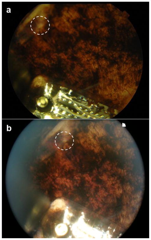
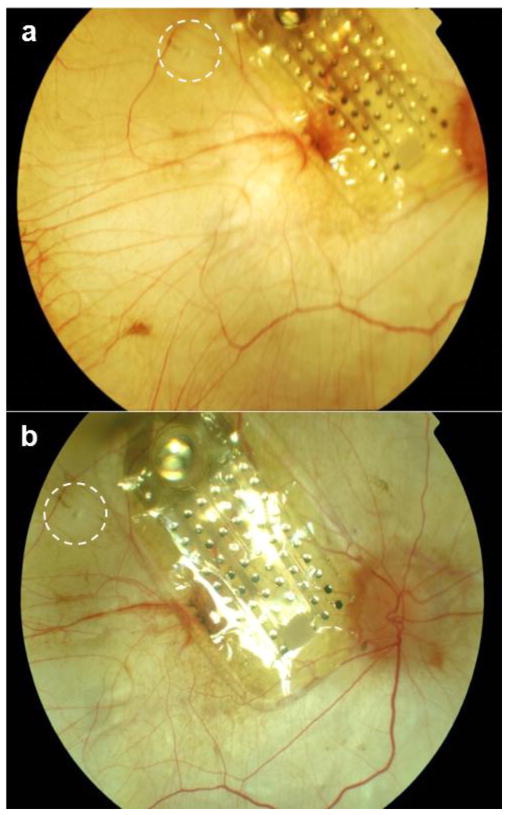
Methods: Custom-built, titanium alloy retinal tacks were mechanically removed from the posterior coats after prolonged implantation (up to 19 months). The resulting wound was characterized by clinical evaluation, fundus photography, and fluorescein angiography while being monitored for stability over time. The wounds were also compared to earlier published reports of the healing response around retinal tacks in human subjects.
Results: Tack extraction was accomplished successfully, without complication, in all four subjects. The wound site was readily identified by pale scar tissue. No change in the wound size or appearance was noted over many months of post-operative observation (up to 22 months after explant). No adverse effects on overall ocular health were detected.
Conclusion: Extraction of retinal tacks from subjects implanted with epiretinal prostheses can be performed without significant complication. The long-term healing response appears to be stable and localized in eyes afflicted with retinitis pigmentosa or choroideremia. There was also minimal, if any, impact on the local circulatory system. These cases suggest that the use of retinal tacks for anchoring epiretinal visual prostheses does not preclude safe repositioning or removal of the device more than a year after implant.
Trial registration: ClinicalTrials.gov NCT00407602.
Conflict of interest statement
Figures


Similar articles
-
Long-term histological and electrophysiological results of an inactive epiretinal electrode array implantation in dogs.Invest Ophthalmol Vis Sci. 1999 Aug;40(9):2073-81. Invest Ophthalmol Vis Sci. 1999. PMID: 10440263
-
Angiographic findings following tack fixation of a wireless epiretinal retina implant device in blind RP patients.Graefes Arch Clin Exp Ophthalmol. 2011 Sep;249(9):1281-6. doi: 10.1007/s00417-011-1653-5. Epub 2011 Apr 5. Graefes Arch Clin Exp Ophthalmol. 2011. PMID: 21465287 Clinical Trial.
-
Implantation and explantation of an active epiretinal visual prosthesis: 2-year follow-up data from the EPIRET3 prospective clinical trial.Eye (Lond). 2012 Apr;26(4):501-9. doi: 10.1038/eye.2012.35. Epub 2012 Mar 16. Eye (Lond). 2012. PMID: 22422033 Free PMC article.
-
Retinal Prostheses and Artificial Vision.Turk J Ophthalmol. 2019 Sep 3;49(4):213-219. doi: 10.4274/tjo.galenos.2019.44270. Turk J Ophthalmol. 2019. PMID: 31486609 Free PMC article. Review.
-
Artificial vision: needs, functioning, and testing of a retinal electronic prosthesis.Prog Brain Res. 2009;175:317-32. doi: 10.1016/S0079-6123(09)17522-2. Prog Brain Res. 2009. PMID: 19660665 Review.
Cited by
-
Retinal Tacks for Complicated Retinal Detachment: Retinal Tacks in the Times of Modern Small-Gauge Vitrectomy.J Ophthalmol. 2022 Mar 31;2022:1968434. doi: 10.1155/2022/1968434. eCollection 2022. J Ophthalmol. 2022. PMID: 35399160 Free PMC article.
-
Assessment of Postoperative Morphologic Retinal Changes by Optical Coherence Tomography in Recipients of an Electronic Retinal Prosthesis Implant.JAMA Ophthalmol. 2019 Mar 1;137(3):272-278. doi: 10.1001/jamaophthalmol.2018.6375. JAMA Ophthalmol. 2019. PMID: 30605209 Free PMC article.
-
Argus II retinal prosthesis malrotation and repositioning with intraoperative optical coherence tomography in a posterior staphyloma.Clin Ophthalmol. 2015 Nov 24;9:2213-6. doi: 10.2147/OPTH.S96570. eCollection 2015. Clin Ophthalmol. 2015. PMID: 26648688 Free PMC article.
References
-
- Ando F, Kondo J. A Plastic Tack for the Treatment of Retinal Detachment With Giant Tear. American J Ophthalmol. 1983;95:260–261. - PubMed
-
- Brown GC. The use of tacks for the repair of complicated retinal detachment. Trans Pa Acad Ophthalmol Otolaryngol. 1987;39:578–582. - PubMed
-
- Humayun MS, Weiland JD, Fujii GY, Greenberg R, Williamson R, Little J, Mech B, Cimmarusti V, Van Boemel G, Dagnelie G, de Juan E. Visual perception in a blind subject with a chronic microelectronic retinal prosthesis. Vis Res. 2003;43:2573–2581. - PubMed
-
- Gerding H, Benner F, Taneri S. Experimental implantation of epiretinal implants (EPI-RET) with an IOL-type receiver. J Neural Eng. 2007;4:S38–S49. - PubMed
-
- Sachs HG, Gabel VP. Retinal replacement - the development of microelectronic retinal prostheses - experience with subretinal implants and new aspects. Graefes Arch Clin Exp Ophthalmol. 2004;242:717–723. - PubMed
Publication types
MeSH terms
Associated data
Grants and funding
LinkOut - more resources
Full Text Sources
Other Literature Sources
Medical
