. 2013 Nov;195(22):5141-50.
doi: 10.1128/JB.00837-13. Epub 2013 Sep 6.

Finely tuned regulation of the aromatic amine degradation pathway in Escherichia coli

Affiliations

Finely tuned regulation of the aromatic amine degradation pathway in Escherichia coli

Ji Zeng et al. J Bacteriol. 2013 Nov.

Abstract

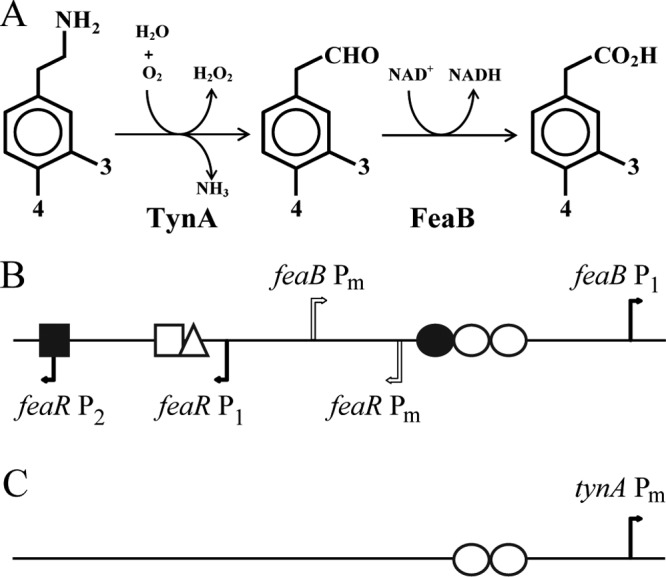
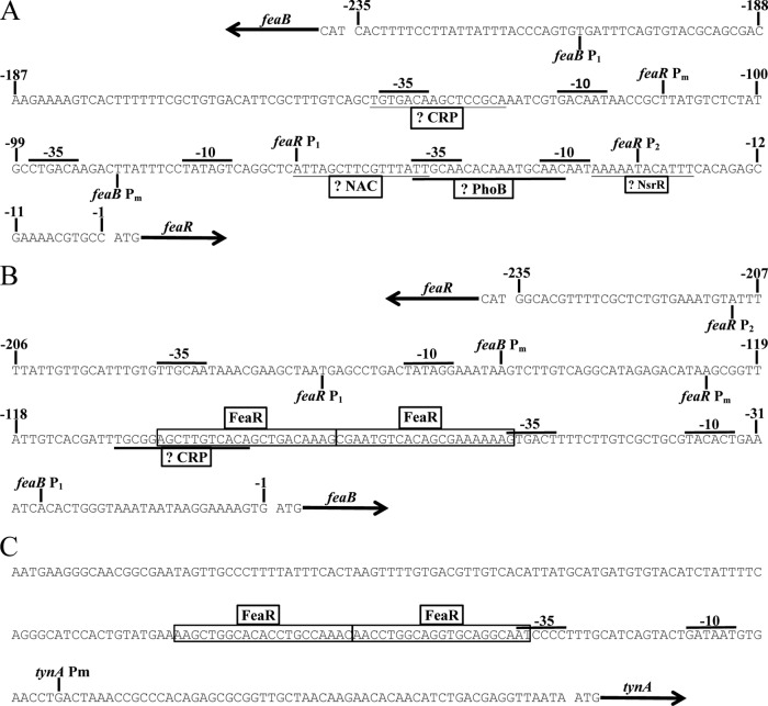
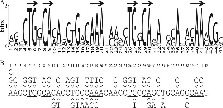
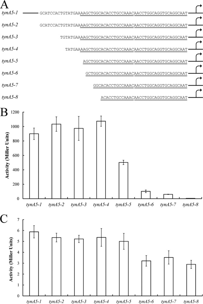
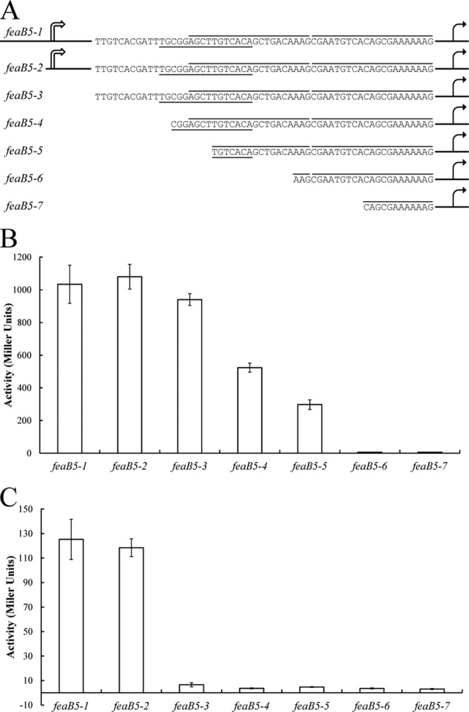
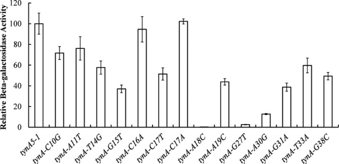
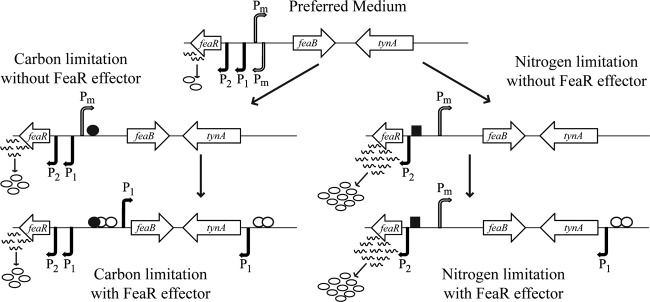
FeaR is an AraC family regulator that activates transcription of the tynA and feaB genes in Escherichia coli. TynA is a periplasmic topaquinone- and copper-containing amine oxidase, and FeaB is a cytosolic NAD-linked aldehyde dehydrogenase. Phenylethylamine, tyramine, and dopamine are oxidized by TynA to the corresponding aldehydes, releasing one equivalent of H2O2 and NH3. The aldehydes can be oxidized to carboxylic acids by FeaB, and (in the case of phenylacetate) can be further degraded to enter central metabolism. Thus, phenylethylamine can be used as a carbon and nitrogen source, while tyramine and dopamine can be used only as sources of nitrogen. Using genetic, biochemical and computational approaches, we show that the FeaR binding site is a TGNCA-N8-AAA motif that occurs in 2 copies in the tynA and feaB promoters. We show that the coactivator for FeaR is the product rather than the substrate of the TynA reaction. The feaR gene is upregulated by carbon or nitrogen limitation, which we propose reflects regulation of feaR by the cyclic AMP receptor protein (CRP) and the nitrogen assimilation control protein (NAC), respectively. In carbon-limited cells grown in the presence of a TynA substrate, tynA and feaB are induced, whereas in nitrogen-limited cells, only the tynA promoter is induced. We propose that tynA and feaB expression is finely tuned to provide the FeaB activity that is required for carbon source utilization and the TynA activity required for nitrogen and carbon source utilization.


Figures

Fig 1
Fig 1
(A) Pathways for the catabolism of phenylethylamine, tyramine, and dopamine. The first reaction is catalyzed by the periplasmic amine oxidase (TynA) and the second reaction by an NAD-linked dehydrogenase (FeaB). Where substituents at the 3 and 4 positions are hydrogen, the three compounds are phenylethylamine, phenylacetaldehyde, and phenylacetate. With a hydroxyl group at the 4 position, they are tyramine, 4-hydroxyphenylacetaldehyde, and 4-hydroxyphenylacetate. With hydroxyl groups at both the 3 and 4 positions, they are dopamine, 3,4-dihydroxyphenylacetaldehyde, and 3,4-dihydroxyphenylacetate. (B and C) Schematics of the organization of the feaR-feaB intergenic region (B) and the tynA regulatory region (C). Transcription start sites are indicated by bent arrows. Verified and predicted binding sites for regulatory proteins are shown: FeaR (open circles), CRP (filled circle), NAC (open triangle), PhoB (open square), and NsrR (filled square). For additional details and DNA sequences, see Fig. 2.
Fig 2
Fig 2
Transcription start sites of feaR (A), feaB (B), and tynA (C) as determined by 5′ RACE. The binding sites for FeaR as defined in this study are boxed. Suggested binding sites for CRP, NAC, PhoB, and NsrR are underlined. Promoter elements (−35 and −10) associated with the mapped transcription start sites are also indicated.
Fig 3
Fig 3
(A) The FeaR binding site. Computational prediction of the FeaR binding site is shown. The tynA and feaB promoter regions from different organisms were used in a search for enriched sequence motifs using the MEME algorithm. Arrows indicate nucleotides that constitute the directly repeated TGNCA-N8-AAA, which is the proposed FeaR consensus binding site. (B) Mutations that were introduced into the FeaR binding site. Nucleotides are numbered according to the sequence logo. Those nucleotides that are within the TGNCA-N8-AAA motif are underlined. Mutations above the sequence were introduced for in vitro DNA binding assays. Mutations below the sequence were used for in vivo reporter fusion assays.
Fig 4
Fig 4
Deletion analysis of the tynA promoters. (A) The tynA promoter was truncated as indicated and fused to lacZ for measurements of β-galactosidase activity. The predicted FeaR binding sites are underlined. (B and C) Promoter activity was measured in cultures grown in PEA medium (B) and in glycerol medium (C). Activities are the means of duplicate measurements from each of three independent cultures, and error bars are standard deviations. The 5′ end of tynA5-1 is shown schematically; this fusion contains 250 bp upstream of the tynA translational start site.
Fig 5
Fig 5
Deletion analysis of the feaB promoters. (A) The feaB promoter was truncated as indicated and fused to lacZ for measurements of β-galactosidase activity. The FeaR binding sites are highlighted by lines above the sequence, and the predicted CRP binding site is underlined. (B and C) Promoter activity was measured in cultures grown in PEA medium (B) and in glycerol medium (C). Activities are the means of duplicate measurements from each of three independent cultures, and error bars are standard deviations. The 5′ ends of feaB5-1 and feaB5-2 are shown schematically; these fusions contain 612 and 250 bp, respectively, upstream of the feaB translational start site.
Fig 6
Fig 6
Gel retardation assay of FeaR CTD binding to the tynA promoter. A fragment from the ytfE promoter was used as a negative control. The labeled DNAs were tynA (lanes 1 to 5) and ytfE (lanes 6 to 10), and the protein concentrations were 0 (lanes 1 and 6), 250 nM (lanes 2 and 7), 500 nM (lanes 3 and 8), 750 nM (lanes 4 and 9), and 1,000 nM (lanes 5 and 10).
Fig 7
Fig 7
(A) Assay of DNA binding by the FeaR CTD by fluorescence anisotropy. The purified CTD was titrated into fluorescently labeled DNAs: site 1 (open squares), site 2 (filled squares), and a negative control, the nrdH promoter (open circles). Each data point is the mean of three measurements, and the plot lines show the fit to equation 1 (see Materials and Methods). The estimated dissociation constants are 301 nM ± 47 nM for site 1 and 219 nM ± 18 nM for site 2. (B) Competition assay using DNAs with mutations in the FeaR binding sites. Unlabeled DNAs were titrated into preformed complexes between the FeaR CTD and a labeled DNA. Each data point is the mean of three determinations, and data were fit to equation 2 (see Materials and Methods). Reactions are shown for DNAs that do not (T5G) (filled squares) and do (C17T) (open squares) compete with the wild-type sequence (competition with wild-type DNA is shown with open circles). Data for all mutations are shown in Table 2.
Fig 8
Fig 8
Site-directed mutagenesis of the FeaR binding site in the tynA promoter (Fig. 3). Mutations were introduced at positions in and near the FeaR binding sites, and mutant promoters were fused to lacZ for measurements of β-galactosidase activity. Activities are the means of duplicate measurements in each of three independent cultures, and error bars are standard deviations.
Fig 9
Fig 9
Summary of the known and proposed mechanisms that regulate transcription of feaR, tynA, and feaB. Active transcription start sites are represented by arrows, with filled arrows for relatively strong promoters and open arrows for weak promoters. The FeaR protein is represented by open circles, CRP-cAMP by filled circles, and NAC by squares. In glycerol-grown cells, feaB is transcribed at low level from Pm, and tynA is not expressed. Upon addition of a FeaR effector, the feaB P1 promoter is activated by FeaR and CRP-cAMP and tynA is activated by FeaR. In cells using glutamine as the nitrogen source, feaB is expressed at a low level from Pm and tynA is not transcribed. In the presence of a FeaR effector, feaB is not expressed, most likely because CRP-cAMP is absent under these conditions. On the other hand, tynA transcription is activated by FeaR. Note that the figure is not drawn to scale.

References

    1. Klinman JP. 2003. The multi-functional topa-quinone copper amine oxidases. Biochim. Biophys. Acta 1647:131–137 - PubMed
    1. Ferrandez A, Prieto MA, Garcia JL, Diaz E. 1997. Molecular characterization of PadA, a phenylacetaldehyde dehydrogenase from Escherichia coli. FEBS Lett. 406:23–27 - PubMed
    1. Diaz E, Ferrandez A, Prieto MA, Garcia JL. 2001. Biodegradation of aromatic compounds by Escherichia coli. Microbiol. Mol. Biol. Rev. 65:523–569 - PMC - PubMed
    1. Teufel R, Mascaraque V, Ismail W, Voss M, Perera J, Eisenreich W, Haehnel W, Fuchs G. 2010. Bacterial phenylalanine and phenylacetate catabolic pathway revealed. Proc. Natl. Acad. Sci. U. S. A. 107:14390–14395 - PMC - PubMed
    1. Hanlon SP, Hill TK, Flavell MA, Stringfellow JM, Cooper RA. 1997. 2-Phenylethylamine catabolism by Escherichia coli K-12: gene organization and expression. J. Gen. Microbiol. 143:513–518 - PubMed

MeSH terms