

The antihelmintic drug pyrvinium pamoate targets aggressive breast cancer
- PMID: 24013655
- PMCID: PMC3754994
- DOI: 10.1371/journal.pone.0071508
The antihelmintic drug pyrvinium pamoate targets aggressive breast cancer
Abstract
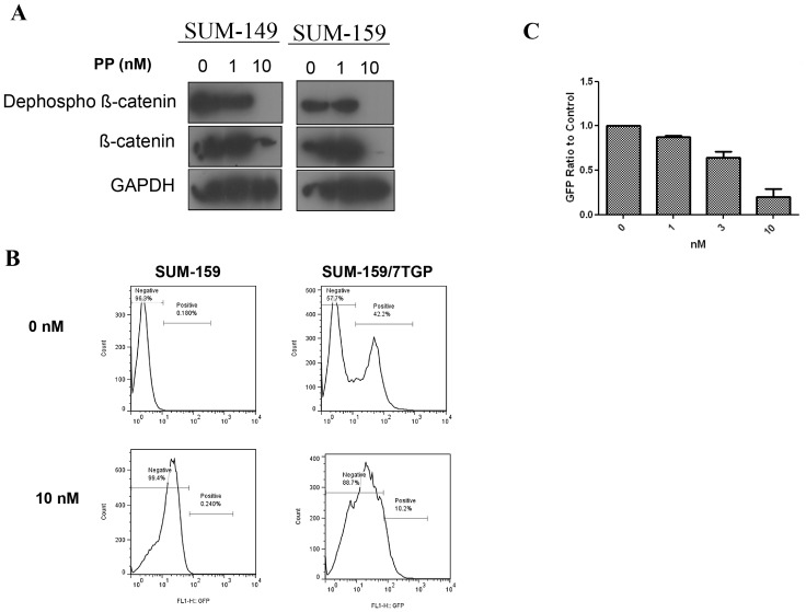
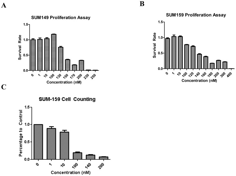
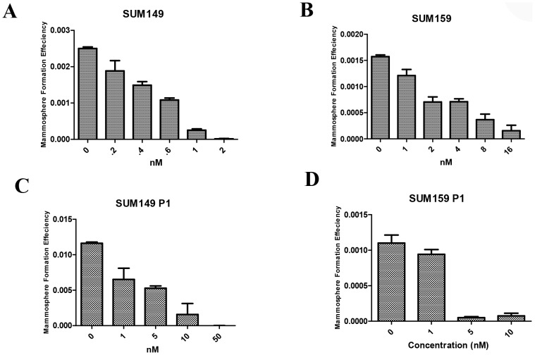
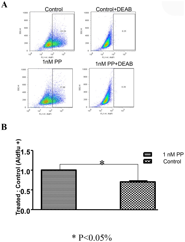
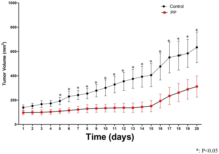
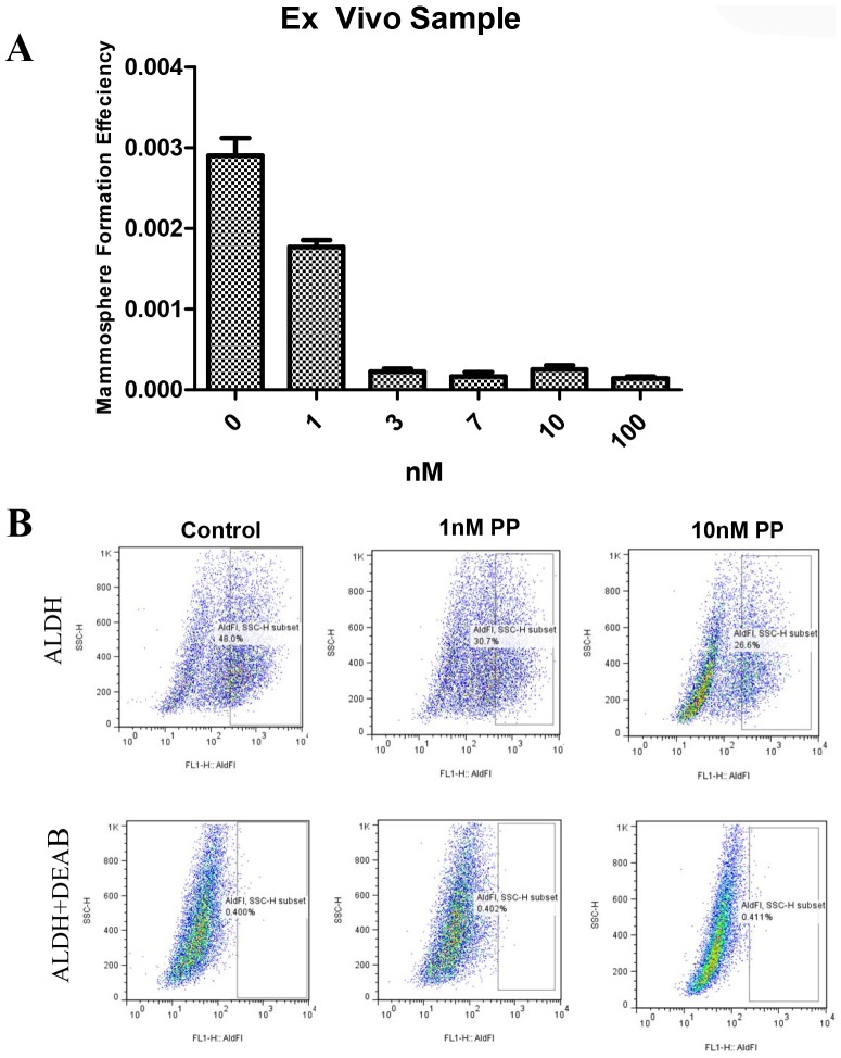
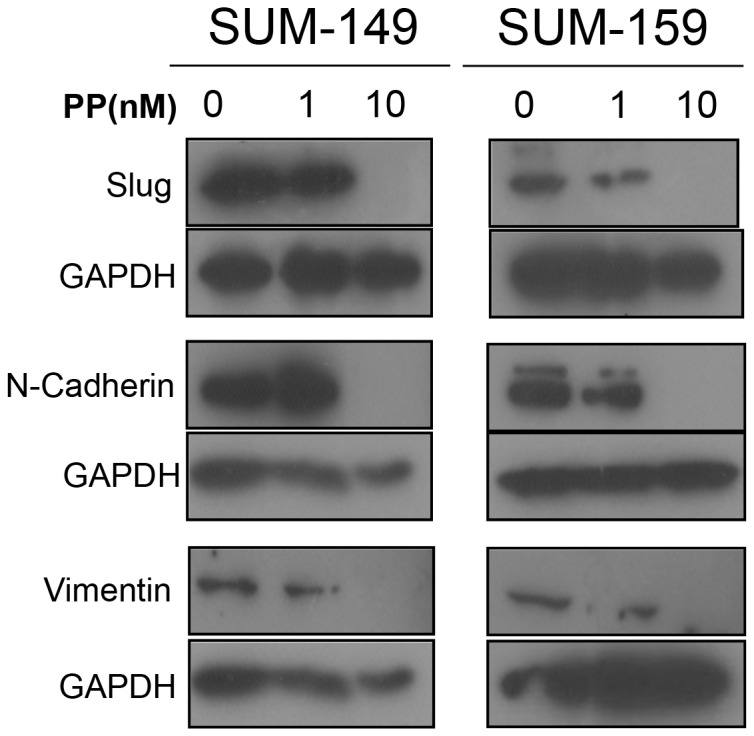
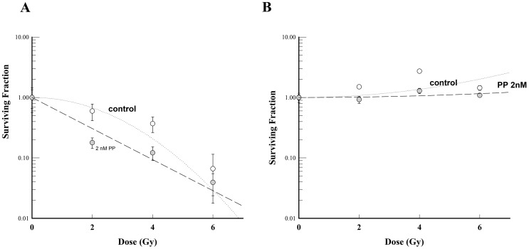
WNT signaling plays a key role in the self-renewal of tumor initiation cells (TICs). In this study, we used pyrvinium pamoate (PP), an FDA-approved antihelmintic drug that inhibits WNT signaling, to test whether pharmacologic inhibition of WNT signaling can specifically target TICs of aggressive breast cancer cells. SUM-149, an inflammatory breast cancer cell line, and SUM-159, a metaplastic basal-type breast cancer cell line, were used in these studies. We found that PP inhibited primary and secondary mammosphere formation of cancer cells at nanomolar concentrations, at least 10 times less than the dose needed to have a toxic effect on cancer cells. A comparable mammosphere formation IC50 dose to that observed in cancer cell lines was obtained using malignant pleural effusion samples from patients with IBC. A decrease in activity of the TIC surrogate aldehyde dehydrogenase was observed in PP-treated cells, and inhibition of WNT signaling by PP was associated with down-regulation of a panel of markers associated with epithelial-mesenchymal transition. In vivo, intratumoral injection was associated with tumor necrosis, and intraperitoneal injection into mice with tumor xenografts caused significant tumor growth delay and a trend toward decreased lung metastasis. In in vitro mammosphere-based and monolayer-based clonogenic assays, we found that PP radiosensitized cells in monolayer culture but not mammosphere culture. These findings suggest WNT signaling inhibition may be a feasible strategy for targeting aggressive breast cancer. Investigation and modification of the bioavailability and toxicity profile of systemic PP are warranted.
Conflict of interest statement
Figures
Similar articles
-
Targeting of Wnt/β-Catenin by Anthelmintic Drug Pyrvinium Enhances Sensitivity of Ovarian Cancer Cells to Chemotherapy.Med Sci Monit. 2017 Jan 16;23:266-275. doi: 10.12659/msm.901667. Med Sci Monit. 2017. PMID: 28090074 Free PMC article.
-
WNT pathway inhibitor pyrvinium pamoate inhibits the self-renewal and metastasis of breast cancer stem cells.Int J Oncol. 2016 Mar;48(3):1175-86. doi: 10.3892/ijo.2016.3337. Epub 2016 Jan 13. Int J Oncol. 2016. PMID: 26781188
-
Inhibitory effect of pyrvinium pamoate on uveal melanoma cells involves blocking of Wnt/β-catenin pathway.Acta Biochim Biophys Sin (Shanghai). 2017 Oct 1;49(10):890-898. doi: 10.1093/abbs/gmx089. Acta Biochim Biophys Sin (Shanghai). 2017. PMID: 28981601
-
The novel role of pyrvinium in cancer therapy.J Cell Physiol. 2018 Apr;233(4):2871-2881. doi: 10.1002/jcp.26006. Epub 2017 Jun 20. J Cell Physiol. 2018. PMID: 28500633 Review.
-
The Wnt/β-catenin pathway in breast cancer therapy: a pre-clinical perspective of its targeting for clinical translation.Expert Rev Anticancer Ther. 2022 Jan;22(1):97-114. doi: 10.1080/14737140.2022.2016398. Epub 2021 Dec 30. Expert Rev Anticancer Ther. 2022. PMID: 34927527 Review.
Cited by
-
Complex HuR function in pancreatic cancer cells.Wiley Interdiscip Rev RNA. 2018 May;9(3):e1469. doi: 10.1002/wrna.1469. Epub 2018 Feb 16. Wiley Interdiscip Rev RNA. 2018. PMID: 29452455 Free PMC article. Review.
-
Targeting of Wnt/β-Catenin by Anthelmintic Drug Pyrvinium Enhances Sensitivity of Ovarian Cancer Cells to Chemotherapy.Med Sci Monit. 2017 Jan 16;23:266-275. doi: 10.12659/msm.901667. Med Sci Monit. 2017. PMID: 28090074 Free PMC article.
-
Discovering gene re-ranking efficiency and conserved gene-gene relationships derived from gene co-expression network analysis on breast cancer data.Sci Rep. 2016 Feb 19;6:20518. doi: 10.1038/srep20518. Sci Rep. 2016. PMID: 26892392 Free PMC article.
-
Pyrvinium pamoate inhibits cell proliferation through ROS-mediated AKT-dependent signaling pathway in colorectal cancer.Med Oncol. 2021 Feb 8;38(2):21. doi: 10.1007/s12032-021-01472-3. Med Oncol. 2021. PMID: 33554313 Free PMC article.
-
Pyrvinium selectively targets blast phase-chronic myeloid leukemia through inhibition of mitochondrial respiration.Oncotarget. 2015 Oct 20;6(32):33769-80. doi: 10.18632/oncotarget.5615. Oncotarget. 2015. PMID: 26378050 Free PMC article.
References
-
- Grudzien P, Lo S, Albain KS, Robinson P, Rajan P, et al. (2010) Inhibition of Notch signaling reduces the stem-like population of breast cancer cells and prevents mammosphere formation. Anticancer Res 30: 3853–3867. - PubMed
Publication types
MeSH terms
Substances
Grants and funding
LinkOut - more resources
Full Text Sources
Other Literature Sources
Medical
Miscellaneous
