. 2013 Sep 6;3(9):e141.
doi: 10.1038/bcj.2013.37.

A novel selective small-molecule PI3K inhibitor is effective against human multiple myeloma in vitro and in vivo

Affiliations

A novel selective small-molecule PI3K inhibitor is effective against human multiple myeloma in vitro and in vivo

J Glauer et al. Blood Cancer J. .

Abstract

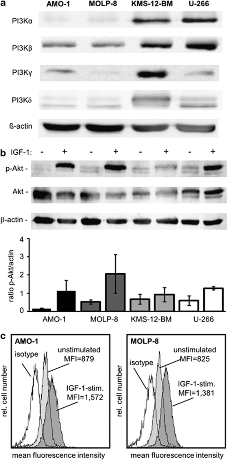
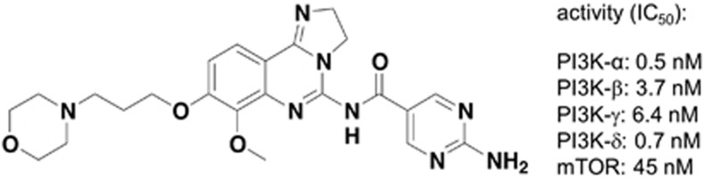
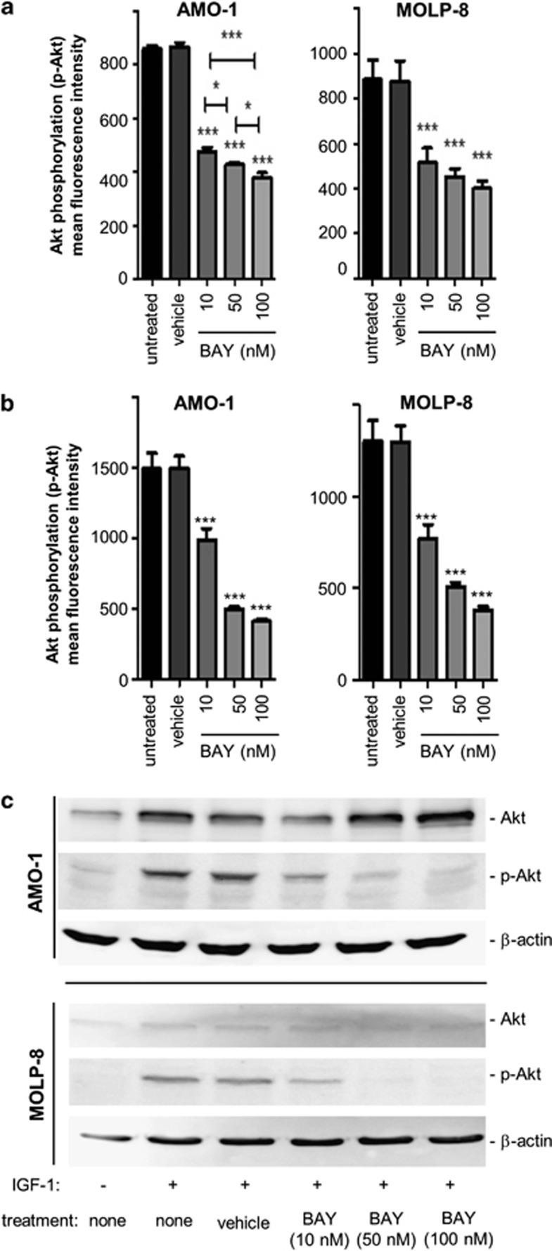
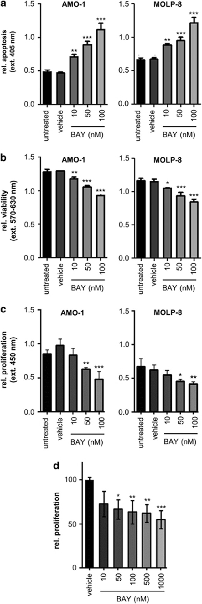
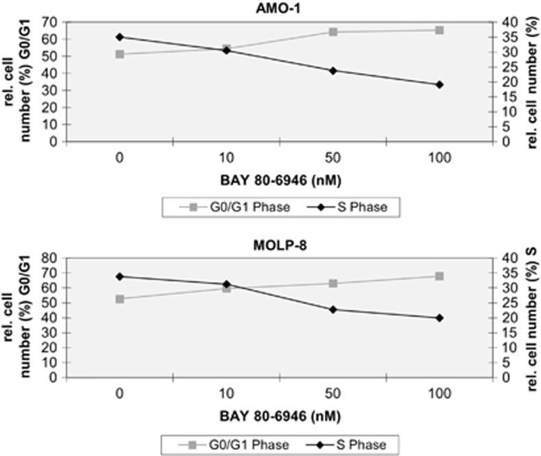
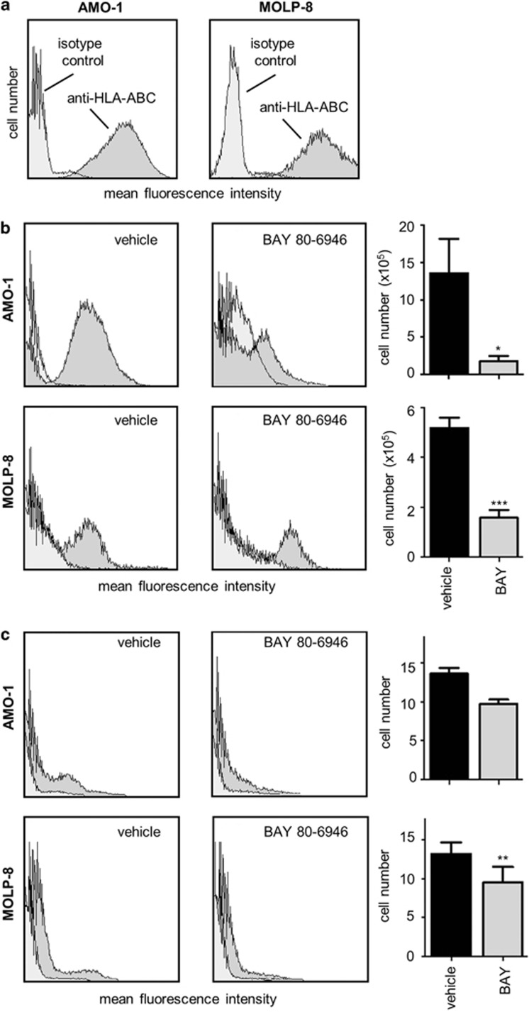
Developing effective therapies against multiple myeloma (MM) is an unresolved challenge. Phosphatidylinositol-3-kinase (PI3K) activation may be associated with tumor progression and drug resistance, and inhibiting PI3K can induce apoptosis in MM cells. Thus, targeting of PI3K is predicted to increase the susceptibility of MM to anticancer therapy. The lead compound of a novel class of PI3K inhibitors, BAY80-6946 (IC50=0.5 nM against PI3K-α), was highly efficacious in four different MM cell lines, where it induced significant antitumoral effects in a dose-dependent manner. The compound inhibited cell cycle progression and increased apoptosis (P<0.001 compared with controls). Moreover, it abrogated the stimulation conferred by insulin-like growth-factor-1, a mechanism relevant for MM progression. These cellular effects were paralleled by decreased Akt phosphorylation, the main downstream target of PI3K. Likewise, profound antitumoral activity was observed ex vivo, as BAY80-6946 significantly inhibited proliferation of freshly isolated myeloma cells from three patients (P<0.001 compared with vehicle). In addition, BAY80-6946 showed convincing in vivo activity against the human AMO-1 and MOLP-8 myeloma cell lines in a preclinical murine xenograft model, where treatment with 6 mg/kg every other day for 2 weeks reduced the cell numbers by 87.0% and 69.3%, respectively (P<0.001 compared with vehicle), without overt toxicity in treated animals.


Figures

Figure 1
Figure 1
Different expression levels of PI3K isoforms and constitutive or IGF-1-stimulated activation of Akt in human myeloma cells. (a) Expression of PI3K isoforms-α, -β, -γ and -δ was examined by western blot analysis in whole-cell lysates of four untreated myeloma cell lines, AMO-1, MOLP-8, KMS-12-BM and U-266. (b) To examine activation of Akt, cellular extracts of native and IGF-1-stimulated cells (100 nM for 20 min) were prepared and western Blot analysis was performed. In the bottom panel, the p-Akt/Akt ratio from two independent experiments was determined densitometrically (± s.e.m). (c) Activation (phosphorylation) of Akt was investigated in two myeloma cell lines either under non-stimulated conditions (light gray histograms) or after stimulation with IGF-1 at a concentration of 100 ng/ml for 20 min. Cells were fixed, permeabilized and stained with an anti-human phospho-Akt antibody and analyzed by flow cytometry.
Figure 2
Figure 2
The novel PI3K inhibitor, BAY 80-6946, inhibits all four PI3K isoforms. Chemical structure of BAY 80-6946 (7-methoxy-8-(3-morpholin-4-ylpropoxy)-2,3-dihydroimidazo [1,2-c]quinazolin-5-amine). The right hand side of the panel depicts IC50 values regarding inhibition of PI3K-α, PI3K-β, PI3K-γ, PI3K-δ and mTOR, respectively, as determined by cell-free assays.
Figure 3
Figure 3
BAY 80-6946 diminishes constitutive and IGF-1-induced phosphorylation of Akt in human myeloma cells. (a) Inhibition of constitutive Akt activation by BAY 80-6946 was examined in the AMO-1 (left) and MOLP-8 (right) human myeloma cell lines. Cells were treated, fixed, permeabilized and stained with an anti-human phosphor-Akt antibody and analyzed by flow cytometry. The values shown represent the averages of three independent experiments (±s.d.). *indicates P<0.05 and ***indicates P<0.001 as compared with vehicle-treated controls. (b) Inhibition of Akt activation by BAY 80-6946 was examined in two IGF-1-stimulated myeloma lines, AMO-1 and MOLP-8. Cells were treated with BAY 80-6946 at the indicated concentrations and then stimulated with 100 ng/ml of IGF-1 for 20 min. Expression of phospho-Akt (p-Akt) was determined by flow cytometry. The values shown represent the average mean fluorescence intensity of three independent experiments (±s.d.). *** indicates P<0.001 as compared with vehicle-treated controls. (c) Inhibition of IGF-1-induced Akt phosphorylation in myeloma cell lines exposed to the indicated concentrations of BAY 80-6946 was confirmed by western blot analysis. The experiment shown is representative of two independent experiments showing similar results.
Figure 4
Figure 4
Induction of apoptosis and decreased viability and proliferation of human myeloma cells by BAY 80-6946 in vitro and ex vivo. (a) The pro-apoptotic activity of BAY 80-6946 was investigated using the Cell Death Detection ELISA Plus measuring the generation of histone-bound DNA fragments. Cells were treated with BAY 80-6946 for 16 h, lysed and incubated with anti-DNA-POD, and anti-histone solution for 2 h. Apoptotic responses were quantitated using a microplate reader at 405 nm. The values shown represent averages (±s.d.) of triplicate measurements from a representative experiment, which was repeated twice with similar results. (b) Viability of human myeloma cells was assessed using an MTT assay. Cells were treated with BAY 80-6946 for 24 h and the number of viable cells was measured using the Cell Titer 96 Aqueous One Solution Cell Proliferation Assay. The values shown represent the averages (±s.d.) of triplicate measurements from a representative experiment, which was repeated twice with similar results. (c) The impact of BAY80-6946 on myeloma cell proliferation was examined by DNA incorporation of the pyrimidine analog BrdU. Myeloma cells were cultured in FCS-free RPMI medium in the presence of BAY80-6946 for 24 h. After 20 h, the cells were stimulated with 100 ng/ml IGF-1 for 20 min. Subsequently, BrdU was added for 4 h and DNA incorporation was determined colorimetrically using the Cell Proliferation ELISA. The values shown represent the average of triplicate measurements (±s.d.) from a representative experiment. The experiment was repeated twice with similar results. (d) Primary human myeloma cells were isolated from blood samples of three patients with high tumor burden. Myeloma cells were ex vivo cultured in RPMI with 0.5% FCS and exposed to BAY80-6946 for 24 h at the indicated concentrations and with 100 ng/ml IGF-1 for 16 h. The values shown represent the average proliferation as determined by BrdU incorporation of myeloma cells from three individual donors (±s.e.m). In all panels, * indicates P<0.05, ** indicates P<0.01, and *** indicates P<0.001, as compared with vehicle-treated controls.
Figure 5
Figure 5
BAY 80-6946 impairs cell cycle progression of human myeloma cells. The influence of BAY 80-6946 on cell cycle progression was examined in two myeloma cell lines, AMO-1 (upper panel) and MOLP-8 (bottom panel). After 24 h of culture in the presence of BAY 80-6946 at the indicated concentrations, the cells were fixed, washed and resuspended with propidium iodide and RNase A, and analyzed by flow cytometry. The graph depicts the relative cell number in G0/G1 and S phase, depending on the concentration of BAY 80-6946.
Figure 6
Figure 6
BAY 80-6946 inhibits myeloma progression in a murine model in vivo. (a) To identify human myeloma cells, cultured AMO-1 and MOLP-8 cells were stained using an antibody directed against HLA-ABC and analyzed by flow cytometry (dark gray histograms). Isotype-matched IgG served as negative control (light gray histograms). (b) To assess the in vivo efficacy of BAY 80-6946, a xenograft model using NU/J Foxn1nu mice transplanted i.p. with 5 × 10⁶ human myeloma cells was used. The experiments were performed using AMO-1 and MOLP-8 cells. Recipient mice were injected i.p. three times per week for 2 weeks with BAY 80-6946 at doses of 6 mg/kg body weight. After the experiment was completed, the total cell number was determined by a thorough diagnostic peritoneal lavage. Peritoneal cells were washed, stained with anti-human HLA-ABC and analyzed by flow cytometry to detect human cells. The histograms exemplify typical findings from representative mice, whereas the graphs depict means (±s.d.) from all animals (n=5 mice per group in MOLP-8 recipients, and n=4 mice per group in AMO-1 recipients). * indicates P<0.05 and *** indicates P<0.001, as compared with vehicle-treated controls. (c) To identify human tumor cells in the spleens of the recipient mice, suspensions of splenocytes were prepared, stained with anti-human HLA-ABC and analyzed by flow cytometry. Again, the histograms exemplify typical findings from representative mice, whereas the graphs depict means (±s.d.) from all animals. ** indicates P<0.01 as compared with vehicle-treated controls.

Similar articles

Cited by

References

    1. Raab MS, Podar K, Breitkreutz I, Richardson PG, Anderson KC. Multiple myeloma. Lancet. 2009;374:324–339. - PubMed
    1. Attal M, Harousseau J-L, Facon T, Guilhot F, Doyen C, Fuzibet JG, et al. Single versus double autologous stem-cell transplantation for multiple myeloma. N Engl J Med. 2003;349:2495–2505. - PubMed
    1. Kyle RA, Rajkumar SV. Multiple myeloma. N Engl J Med. 2004;351:1860–1873. - PubMed
    1. Lenhoff S, Hjorth M, Holmberg E, Turesson I, Westin J, Nielsen JL, et al. Impact on survival of high-dose therapy with autologous stem cell support in patients younger than 60 years with newly diagnosed multiple myeloma: a population-based study. Nordic Myeloma Study Group. Blood. 2000;95:7–11. - PubMed
    1. Richardson P, Hideshima T, Anderson KC. An update of novel therapeutic approaches for multiple myeloma. Curr Treat Options Oncol. 2004;5:227–238. - PubMed