

Region specific response of intervertebral disc cells to complex dynamic loading: an organ culture study using a dynamic torsion-compression bioreactor
- PMID: 24013824
- PMCID: PMC3755972
- DOI: 10.1371/journal.pone.0072489
Region specific response of intervertebral disc cells to complex dynamic loading: an organ culture study using a dynamic torsion-compression bioreactor
Abstract
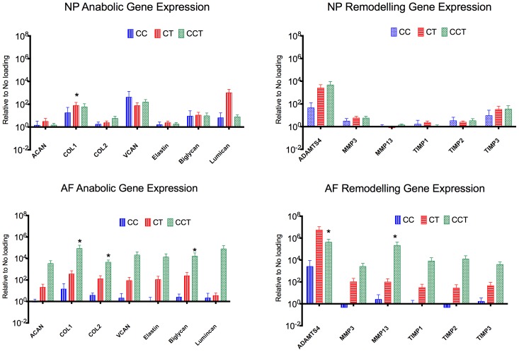
The spine is routinely subjected to repetitive complex loading consisting of axial compression, torsion, flexion and extension. Mechanical loading is one of the important causes of spinal diseases, including disc herniation and disc degeneration. It is known that static and dynamic compression can lead to progressive disc degeneration, but little is known about the mechanobiology of the disc subjected to combined dynamic compression and torsion. Therefore, the purpose of this study was to compare the mechanobiology of the intervertebral disc when subjected to combined dynamic compression and axial torsion or pure dynamic compression or axial torsion using organ culture. We applied four different loading modalities [1. control: no loading (NL), 2. cyclic compression (CC), 3. cyclic torsion (CT), and 4. combined cyclic compression and torsion (CCT)] on bovine caudal disc explants using our custom made dynamic loading bioreactor for disc organ culture. Loads were applied for 8 h/day and continued for 14 days, all at a physiological magnitude and frequency. Our results provided strong evidence that complex loading induced a stronger degree of disc degeneration compared to one degree of freedom loading. In the CCT group, less than 10% nucleus pulposus (NP) cells survived the 14 days of loading, while cell viabilities were maintained above 70% in the NP of all the other three groups and in the annulus fibrosus (AF) of all the groups. Gene expression analysis revealed a strong up-regulation in matrix genes and matrix remodeling genes in the AF of the CCT group. Cell apoptotic activity and glycosaminoglycan content were also quantified but there were no statistically significant differences found. Cell morphology in the NP of the CCT was changed, as shown by histological evaluation. Our results stress the importance of complex loading on the initiation and progression of disc degeneration.
Conflict of interest statement
Figures


Similar articles
-
Functional impact of integrin α5β1 on the homeostasis of intervertebral discs: a study of mechanotransduction pathways using a novel dynamic loading organ culture system.Spine J. 2015 Mar 1;15(3):417-26. doi: 10.1016/j.spinee.2014.12.143. Epub 2014 Dec 27. Spine J. 2015. PMID: 25546513 Free PMC article.
-
Duration-dependent influence of dynamic torsion on the intervertebral disc: an intact disc organ culture study.Eur Spine J. 2015 Nov;24(11):2402-10. doi: 10.1007/s00586-015-4140-6. Epub 2015 Jul 28. Eur Spine J. 2015. PMID: 26215177
-
A repeated strike loading organ culture model for studying compression-associated chronic disc degeneration.Biomol Biomed. 2025 Jan 30;25(3):708-719. doi: 10.17305/bb.2024.10640. Biomol Biomed. 2025. PMID: 39101754 Free PMC article.
-
Mechanobiology of the intervertebral disc and relevance to disc degeneration.J Bone Joint Surg Am. 2006 Apr;88 Suppl 2:52-7. doi: 10.2106/JBJS.F.00001. J Bone Joint Surg Am. 2006. PMID: 16595444 Review.
-
The Influence of Axial Compression on the Cellular and Mechanical Function of Spinal Tissues; Emphasis on the Nucleus Pulposus and Annulus Fibrosus: A Review.J Biomech Eng. 2021 May 1;143(5):050802. doi: 10.1115/1.4049749. J Biomech Eng. 2021. PMID: 33454730 Review.
Cited by
-
Intervertebral Disc-on-a-Chip as Advanced In Vitro Model for Mechanobiology Research and Drug Testing: A Review and Perspective.Front Bioeng Biotechnol. 2022 Jan 28;9:826867. doi: 10.3389/fbioe.2021.826867. eCollection 2021. Front Bioeng Biotechnol. 2022. PMID: 35155416 Free PMC article. Review.
-
The role of biomechanical factors in models of intervertebral disc degeneration across multiple length scales.APL Bioeng. 2023 May 8;7(2):021501. doi: 10.1063/5.0137698. eCollection 2023 Jun. APL Bioeng. 2023. PMID: 37180733 Free PMC article. Review.
-
Developments in intervertebral disc disease research: pathophysiology, mechanobiology, and therapeutics.Curr Rev Musculoskelet Med. 2015 Mar;8(1):18-31. doi: 10.1007/s12178-014-9253-8. Curr Rev Musculoskelet Med. 2015. PMID: 25694233 Free PMC article.
-
Effect of Static Load on the Nucleus Pulposus of Rabbit Intervertebral Disc Motion Segment in Ex vivo Organ Culture.Chin Med J (Engl). 2016 Oct 5;129(19):2338-46. doi: 10.4103/0366-6999.190666. Chin Med J (Engl). 2016. PMID: 27647194 Free PMC article.
-
The influence of intervertebral disc overloading on nociceptor calcium flickering.JOR Spine. 2023 Jun 9;6(3):e1267. doi: 10.1002/jsp2.1267. eCollection 2023 Sep. JOR Spine. 2023. PMID: 37780827 Free PMC article.
References
-
- Elliott B, Khangure M (2002) Disk degeneration and fast bowling in cricket: an intervention study. Med Sci Sports Exerc 34: 1714–1718. - PubMed
-
- Grimshaw P, Giles A, Tong R, Grimmer K (2002) Lower back and elbow injuries in golf. Sports Med 32: 655–666. - PubMed
-
- Hangai M, Kaneoka K, Hinotsu S, Shimizu K, Okubo Y, et al. (2009) Lumbar intervertebral disk degeneration in athletes. Am J Sports Med 37: 149–155. - PubMed
-
- Norman R, Wells R, Neumann P, Frank J, Shannon H, et al. (1998) A comparison of peak vs cumulative physical work exposure risk factors for the reporting of low back pain in the automotive industry. Clin Biomech (Bristol, Avon) 13: 561–573. - PubMed
-
- Kelsey JL, Githens PB, White AA, Holford TR, Walter SD, et al. (1984) An epidemiologic study of lifting and twisting on the job and risk for acute prolapsed lumbar intervertebral disc. J Orthop Res 2: 61–66. - PubMed
Publication types
MeSH terms
Substances
LinkOut - more resources
Full Text Sources
Other Literature Sources
Miscellaneous
