. 2013 Aug 27;8(8):e73209.
doi: 10.1371/journal.pone.0073209. eCollection 2013.

Circadian clock regulation of the cell cycle in the zebrafish intestine

Affiliations

Circadian clock regulation of the cell cycle in the zebrafish intestine

Elodie Peyric et al. PLoS One. .

Abstract

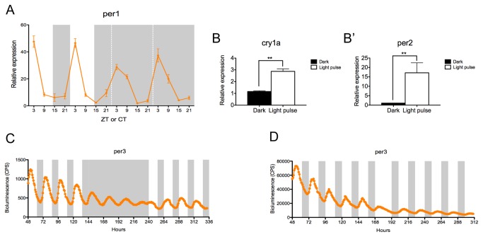
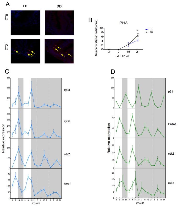
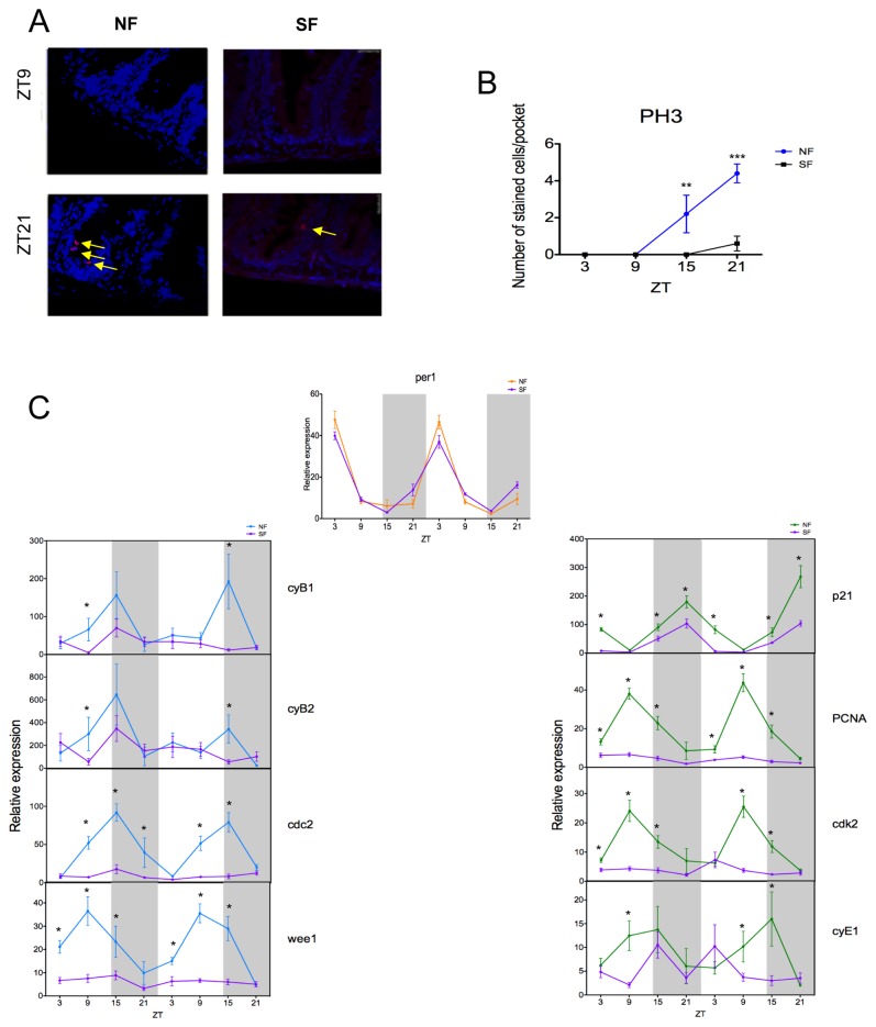
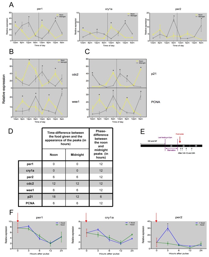
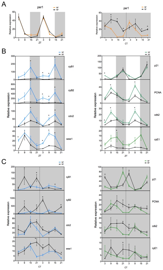
The circadian clock controls cell proliferation in a number of healthy tissues where cell renewal and regeneration are critical for normal physiological function. The intestine is an organ that typically undergoes regular cycles of cell division, differentiation and apoptosis as part of its role in digestion and nutrient absorption. The aim of this study was to explore circadian clock regulation of cell proliferation and cell cycle gene expression in the zebrafish intestine. Here we show that the zebrafish gut contains a directly light-entrainable circadian pacemaker, which regulates the daily timing of mitosis. Furthermore, this intestinal clock controls the expression of key cell cycle regulators, such as cdc2, wee1, p21, PCNA and cdk2, but only weakly influences cyclin B1, cyclin B2 and cyclin E1 expression. Interestingly, food deprivation has little impact on circadian clock function in the gut, but dramatically reduces cell proliferation, as well as cell cycle gene expression in this tissue. Timed feeding under constant dark conditions is able to drive rhythmic expression not only of circadian clock genes, but also of several cell cycle genes, suggesting that food can entrain the clock, as well as the cell cycle in the intestine. Rather surprisingly, we found that timed feeding is critical for high amplitude rhythms in cell cycle gene expression, even when zebrafish are maintained on a light-dark cycle. Together these results suggest that the intestinal clock integrates multiple rhythmic cues, including light and food, to function optimally.


Conflict of interest statement

Competing Interests: The authors have declared that no competing interests exist.

Figures

Figure 1
Figure 1. Zebrafish intestine possesses a directly light-responsive circadian pacemaker.
(A) After entrainment to a LD cycle (14L:10D), the expression of the core clock component per1 is rhythmic in the intestine with a peak at ZT3. The oscillation is maintained when animals free-run in DD. Data represents the mean ± SEM from 8 fish per indicated zeitgeber or circadian time (ZT or CT), where ZT0 is lights on. (B–B’) A three-hour light pulse induces expression of cry1a and per2 compared to a dark control. Data represent the mean ± SEM from 5 fish. (C) The adult intestine of per3-luciferase zebrafish entrained to 4 days of LD, 4 days of DD and returned to 4 days in LD was monitored. Gut per3 expression is rhythmic in LD with a peak at ZT5 and free-runs in DD with a damped amplitude. The mean bioluminescence in counts per seconds (CPS) is plotted (n=3-4). (D) Intestine of adult per3-luciferase zebrafish were entrained to 5 days of LD then transferred in DL for 6 days. Gut per3 is able to re-entrain to a new, reversed light regime. The mean bioluminescence in CPS is plotted (n=3-4). White and grey backgrounds represent light and dark phases, respectively.
Figure 2
Figure 2. M phase is rhythmic and under circadian control.
(A) In LD and DD conditions, cells stained with an anti-pH3 antibody to monitor mitosis (in red and marked by the yellow arrow) at the peak and trough time points, are localized inside the intervillus pockets of the gut. (B) Cell division is rhythmic and is exhibiting a peak of stained cells per pocket at ZT21. This rhythmicity is maintained in DD, showing that cell division is under circadian control. DAPI is used here as a nuclear counterstain. (C–D) Quantitative PCR analysis of endogenous cell cycle genes from M phase (C) and G1/S phase (D). (C) In LD, cyB1, cyB2 and cdc2 are rhythmic and peak at ZT15, whereas wee1 peaks at ZT9. All genes continue to oscillate after entry into DD. (D) In LD, p21, PCNA and cdk2 show a strong rhythm with peak expression at ZT9 for PCNA and cdk2 and a peak at ZT21 for p21. In DD, all genes continue to oscillate robustly with the possible exception of cyE1, which shows quite variable expression even in LD. White and grey backgrounds represent light and dark phases, respectively. Cell cycle gene expression data represents the mean ± SEM from 8 fish per time point. For each time point in panel B, LD data are compared to DD using a Student’s t-test (* p<0.05). All other data are expressed as mean ± SEM.
Figure 3
Figure 3. M phase is affected by starvation.
(A) Cell division is rhythmic under a normal feeding schedule (NF) (peak and trough shown), but this rhythm is lost when fish are starved (SF). DAPI is used here as a nuclear counterstain. (B) Cell division is largely abolished when no food is given. (C) The per1 rhythm is unaltered in NF and SF fish. However, all the M-phase genes studied (cyB1, cyB2, cdc2 and wee1) and most G1/S-phase genes (PCNA, cdk2 and cyE1) show reduced levels of expression, and a general loss of rhythmicity during starvation. p21 expression is the one exception, showing a relatively small response to starvation. White and grey backgrounds represent light and dark phases, respectively. Cell cycle gene data represents the mean ± SEM from 8 to 12 fish per time point. For panels B and C, NF data are compared to SF using a Student’s t-test (* p<0.05, ** p<0.01 and *** p<0.001).
Figure 4
Figure 4. Intestinal circadian clock and cell cycle genes are food-entrainable in zebrafish.
(A) In DD, clock genes per1, cry1a and per2 are rhythmically expressed during restricted feeding, when food is provided at noon or midnight. The rhythms in clock gene expression retain a stable phase relationship between the two opposite feeding schedules. (B–C) Key cell cycle regulators show corresponding entrainment to the two opposite feeding regimes. cdc2 peaks are observed at midnight for noon fed animals and at midday for the midnight fed animals. wee1 expression shows peak values 6 hours after the feeding time. (D) The table illustrates the time difference between the feeding regime, noon or midnight, and peak expression for all the genes studied, as well as the phase difference between the two experiments. (E) A schematic of the food pulse experimental design. (F) There is no acute effect of feeding for per1 and cry1a expression. In contrast, per2 expression after 3h is increased compared to the unfed control. Red arrow represents timing of the food pulse. Grey backgrounds represent constant dark conditions. Data represents the mean ± SEM from 3 or 4 fish per time point. For panels A, B and C, samples collected at noon are compared to those collected at midnight using a Student’s t-test. For panel D, at each time point after the food pulse, a Student’s t-test is employed to compare the two conditions (* represents a significant statistical difference of p<0.05).
Figure 5
Figure 5. Random feeding in LD and DD alters cell cycle gene oscillations.
(A) Entrained to a LD cycle, the rhythm and amplitude of per1 expression is not altered by the feeding regime, either normal (NF) or random (RF). In DD and NF, per1 is rhythmic, but under RF and DD conditions, per1 ceases to show a robust, precise daily rhythm. (B) Cyclin gene expression (cyB1, cyB2 and cyE1) shows a large reduction in the level of expression and very shallow oscillations in RF compared to NF. Under RF, expression of cdc2, wee1, PCNA and cdk2 displays a similar rhythmicity to NF, but with significantly reduced amplitude. p21 expression is not altered by the feeding regime. (C) When fish free-run in DD and are fed at random times, all the genes studied (cyB1, cyB2, cdc2, wee1, PCNA, cdk2 and cyE1), except p21, display a disrupted profile compared to NF. No clear rhythmicity is observed. White and grey backgrounds represent light and dark phases, respectively. Uninterrupted grey backgrounds represent constant dark condition. Data represents the mean ± SEM from 3 or 4 fish per time point. For panels A, B and C, NF data are compared to RF at each time point using a Student’s t-test (* represents a significant statistical difference of p<0.05).

Similar articles

Cited by

References

    1. Reppert SM, Weaver DR (2002) Coordination of circadian timing in mammals. Nature 418: 935-941. doi:10.1038/nature00965. PubMed: 12198538. - DOI - PubMed
    1. Panda S, Hogenesch JB, Kay SA (2002) Circadian rhythms from flies to human. Nature 417: 329-335. doi:10.1038/417329a. PubMed: 12015613. - DOI - PubMed
    1. Bell-Pedersen D, Cassone VM, Earnest DJ, Golden SS, Hardin PE et al. (2005) Circadian rhythms from multiple oscillators: lessons from diverse organisms. Nat Rev Genet 6: 544-556. doi:10.1038/nrg1633. PubMed: 15951747. - DOI - PMC - PubMed
    1. Hoogerwerf WA (2006) Biologic clocks and the gut. Curr Gastroenterol Rep 8: 353-359. doi:10.1007/s11894-006-0019-3. PubMed: 16968601. - DOI - PubMed
    1. Konturek PC, Brzozowski T, Konturek SJ (2011) Gut clock: implication of circadian rhythms in the gastrointestinal tract. J Physiol Pharmacol 62: 139-150. PubMed: 21673361. - PubMed

Publication types

MeSH terms

LinkOut - more resources