. 2013 Oct 18;288(42):30192-30209.
doi: 10.1074/jbc.M113.503060. Epub 2013 Sep 6.

O-Linked β-N-acetylglucosamine (O-GlcNAc) regulates emerin binding to barrier to autointegration factor (BAF) in a chromatin- and lamin B-enriched "niche"

Affiliations

O-Linked β-N-acetylglucosamine (O-GlcNAc) regulates emerin binding to barrier to autointegration factor (BAF) in a chromatin- and lamin B-enriched "niche"

Jason M Berk et al. J Biol Chem. .

Abstract

Emerin, a membrane component of nuclear "lamina" networks with lamins and barrier to autointegration factor (BAF), is highly O-GlcNAc-modified ("O-GlcNAcylated") in mammalian cells. Mass spectrometry analysis revealed eight sites of O-GlcNAcylation, including Ser-53, Ser-54, Ser-87, Ser-171, and Ser-173. Emerin O-GlcNAcylation was reduced ~50% by S53A or S54A mutation in vitro and in vivo. O-GlcNAcylation was reduced ~66% by the triple S52A/S53A/S54A mutant, and S173A reduced O-GlcNAcylation of the S52A/S53A/S54A mutant by ~30%, in vivo. We separated two populations of emerin, A-type lamins and BAF; one population solubilized easily, and the other required sonication and included histones and B-type lamins. Emerin and BAF associated only in histone- and lamin-B-containing fractions. The S173D mutation specifically and selectively reduced GFP-emerin association with BAF by 58% and also increased GFP-emerin hyper-phosphorylation. We conclude that β-N-acetylglucosaminyltransferase, an essential enzyme, controls two regions in emerin. The first region, defined by residues Ser-53 and Ser-54, flanks the LEM domain. O-GlcNAc modification at Ser-173, in the second region, is proposed to promote emerin association with BAF in the chromatin/lamin B "niche." These results reveal direct control of a conserved LEM domain nuclear lamina component by β-N-acetylglucosaminyltransferase, a nutrient sensor that regulates cell stress responses, mitosis, and epigenetics.

Keywords: Cardiomyopathy; Chromosomes/Non-histone Chromosomal Proteins; Emerin; Lamin; Laminopathy; Muscular Dystrophy; Nuclear Matrix; Nuclear Membrane; Nucleoskeleton; O-GlcNAc.


Figures
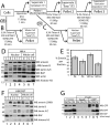
FIGURE 1.
FIGURE 1.
Distribution of endogenous nuclear proteins in unsonicated versus sonicated lysates from HeLa or HEK293T cells (“NE/NS partitioning”). A–C, schematic diagrams of cell fractionation protocols used to isolate the easy versus sonication-dependent fractions of cells or pelleted nuclei (A). Alternatively, we directly lysed and sonicated whole cells (B) or pelleted nuclei (C) to generate total lysates. D and E, equal percentages (2%) of lysates from isolated nuclei or whole HeLa cell lysates (C, cytoplasm; E, easily extracted; S, sonicated; T, total) were resolved by SDS-PAGE and immunoblotted for endogenous cytoplasmic and nuclear proteins. *, B-type lamins; two nonspecific bands were also detected by the lamin B antibody in HeLa cells. The emerin distribution from D was quantified in E as the percentage of emerin in the easy versus sonication-dependent fractions (n = 4; error bars, S.E.). F, same experiment as A for HEK293T cells (1% of fractions; n = 3). G, GFP-emerin and endogenous BAF are present in both the NE and NS fractions of HEK293T cells but associate (co-immunoprecipitate) only in NS. Easy, sonicated, and total nuclear fractions (NE, NS, and NT, respectively) from HEK293T cells 24 h posttransfection with GFP-emerin (wild type or mutant; control lanes show input lysates (1%)) immunoprecipitated with bead-conjugated GFP antibodies or beads alone, resolved by SDS-PAGE, and blotted with antibodies to GFP (WB: GFP) or BAF (WB: BAF; bottom panel shows shorter exposure). Each blot shown is a representative of three independent experiments (n = 3).
FIGURE 2.
FIGURE 2.
Endogenous cellular emerin is O-GlcNAcylated. A, Western blots of unsonicated whole cell lysates from HeLa cells treated with or without the OGA inhibitor TMG (1 μm, 18 h), either before (input, 10 μg) or after immunoprecipitating with nonspecific (IgG) or emerin-specific (αEmr) rabbit antibodies (100% loaded). Duplicate blots were probed first with either O-GlcNAc antibody (CTD110.6) alone (left) or CTD110.6 plus competing 100 mm GlcNAc (right). The position of emerin is boxed. Each blot was stripped and reprobed with the emerin-specific mouse NCL antibody (A, bottom, n = 3). B, Western blots of endogenous emerin immunoprecipitated from equal volumes of HeLa cell NE and NS fractions (1% input shown), resolved by SDS-PAGE, and probed first with the O-GlcNAc antibody (CTD110.6) and then stripped and reprobed for emerin (n = 2). C and D, control versus emerin-specific immunoprecipitates from GFP control cells (GFP) and OGT KO MEFs (OGT). MEFs were immunoblotted using either the O-GlcNAc antibody (CTD110.6) or CTD110.6 plus competing 100 mm GlcNAc and quantified in D as the O-GlcNAc/emerin ratio normalized to each corresponding vehicle (ethanol; Et) control. Error bars, S.E. (n = 3; **, p < 0.004; Student's t test). E, to verify OGT protein reduction, whole cell protein lysates (10 μg/lane) from 4-hydroxytamoxifen (4OHT)-treated (or vehicle control) MEFs (GFP versus OGT KO) were resolved and immunoblotted with antibodies specific for OGT, O-GlcNAc, or α-tubulin.
FIGURE 3.
FIGURE 3.
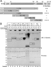
In vitro O-GlcNAc modification of recombinant emerin. A, schematic of full-length emerin (wild type; WT) and tested polypeptides: His- and S-tagged human emerin residues 1–70 or 1–176 and GST-tagged human emerin residues 1–222, 70–140, 140–176, or 170–220. In vitro O-GlcNAcylation results for each polypeptide (B) are summarized qualitatively (+/−) on the right in A. B, in vitro O-GlcNAcylation results. Each purified emerin polypeptide (∼1 μg) was incubated with or without 1 μg of OGT, with or without excess UDP-GlcNAc. Membranes were directly Ponceau-stained to detect proteins (bottom) and then probed with O-GlcNAc-specific antibody CTD110.6 (WB: O-GlcNAc; n = 3). Boxes indicate O-GlcNAc signals for each OGT-treated versus untreated polypeptide. Arrowhead, OGT. One polypeptide (GST 70–140) gave high background signals that did not reproducibly increase in the presence of OGT.
FIGURE 4.
FIGURE 4.
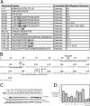
Identification and validation of O-GlcNAc sites in recombinant emerin. A, summary of mass spectrometric analysis of O-GlcNAcylated His-emerin peptides. All 10 Ser/Thr-containing proteolytic peptides were detected. All O-GlcNAc-modified peptides were detected with a mass accuracy error of <5 ppm. Five sites (Ser-53, Ser-54, Ser-87, Ser-171, and Ser-173) were unambiguously assigned. Modified residues are shown in boldface type. *, the third O-GlcNAc site on the tri-O-GlcNAcylated peptide could not be assigned precisely; ETD spectra were consistent with a third O-GlcNAc on Ser-57 in the context of the tri-O-GlcNAcylated peptide but did not rule out potential modification at Ser-58. **, mono- and di-O-GlcNAcylated forms of SSLDLSYYPTSSSTSFMSSSSSSSSWLTR were detected by elution profile, accurate mass, and CAD signature ions. B, amino acid sequence of human emerin residues 1–220, indicating the five identified O-GlcNAc sites. Underlines, O-GlcNAcylated peptides with identified sites; overline, O-GlcNAcylated peptide with at least two unidentified sites. C and D, site validation by [3H]UDP-GlcNAc incorporation into purified missense-mutated emerin in vitro. Shown are a representative Coomassie-stained SDS-PAGE gel (C, bottom) and corresponding autoradiograph (C, top) of [3H]GlcNAc incorporated into recombinant purified His-emerin 1–220 after incubation with (or without) OGT and 1 μCi of [3H]UDP-GlcNAc diluted with unradiolabeled UDP-GlcNAc (final concentration, 8.33 μm; n = 4 or 5 for all mutants except the quintuple S52A/S53A/S54A/S171A/S173A (n = 2) and double S171A/S173A (n = 1) mutants). Results from C were quantified by densitometry and graphed in D as the ratio of the [3H]GlcNAc to Coomassie emerin signals, normalized to that of wild type emerin 1–220. Error bars, S.E.; *, p < 0.02 (Mann-Whitney U test; n = 4 or 5). ND, not detected.
FIGURE 5.
FIGURE 5.
Residues Ser-53, Ser-54, and Ser-173 are relevant to emerin O-GlcNAcylation in HeLa cells. A, direct GFP fluorescence images of HeLa cells 24 h after transfection with GFP-emerin (wild type or missense mutation), showing that mutations do not grossly mislocalize GFP-emerin. DNA was DAPI-stained. Bar, 10 μm. Images were contrast-adjusted using Adobe Photoshop. B and C, immunoblots of unsonicated whole cell protein lysates from HeLa cells (WCLE) that transiently expressed GFP or GFP-emerin (wild type or missense-mutated) for 24 h, resolved by SDS-PAGE either before (1% input) or after immunoprecipitation with rabbit GFP antibodies. Immunoblots were probed first with the O-GlcNAc antibody (WB: O-GlcNAc; top) and then stripped and reprobed for GFP (WB: GFP; bottom). A vertical black line separates different parts of the same blot. C, results from B quantified by densitometry and graphed as the O-GlcNAc-to-GFP-emerin ratio for each mutant, normalized to wild type GFP-emerin. Error bars, S.E.; significance was determined by Student's t test. **, p < 0.007; *, p < 0.04 (n = 3–5). D and E, similar to B, but HeLa cells were treated (or not) for 4 h with 1 μm TMG (which inhibits OGA) to increase O-GlcNAc signals prior to harvest, and whole cell lysates were sonicated (WCLT) prior to immunoprecipitation and immunoblotting first with antibodies to O-GlcNAc (D, top) and then GFP antibodies (D, bottom). Inset, contrast-enhanced (backlit scanned), close-up view of boxed region. E, results for each quadruple mutant (S52–54A plus S171A or S173A) graphed relative to the triple mutant S52–54A. Error bars, S.E.; n = 3; *, p < 0.04 by Student's t test).
FIGURE 6.
FIGURE 6.
S173D mutation disrupts GFP-emerin association with BAF. A, the NE versus NS distributions of GFP or GFP-emerin (wild type, S53A, S54F, S173A, or S173D) were tested in HeLa cells 24 h posttransfection. Shown are equal percentages (2%) of each cytoplasm (C) and separated NE and NS fractions, resolved by SDS-PAGE and immunoblotted with antibodies to emerin, lamin B, lamins A/C, BAF, actin, or histone H3 (n = 3). B and C, sonicated total nuclear lysates (NT) from HEK293T cells 24 h posttransfection with GFP-emerin (wild type or mutant), precipitated with bead-conjugated GFP antibodies or beads alone, SDS-PAGE-resolved, and probed with antibodies to GFP, lamins A/C, or BAF. Controls show input NT lysate (1%) and beads-only precipitation from wild type GFP-emerin lysates. Results are quantified in C as the amount of BAF that co-immunoprecipitated with each GFP-emerin mutant, relative to wild type GFP-emerin (n = 7; **, p < 0.0002 by Student's t test; bars, S.E.).
FIGURE 7.
FIGURE 7.
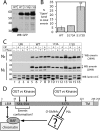
Posttranslational consequences of GFP-emerin S173D versus S173A mutations in HeLa cells and model. A and B, immunoblot of equal protein concentrations of easy whole cell lysates (WCLE) from HeLa cells 24 h posttransfection with GFP or GFP-emerin (wild type, 173A, or 173D), probed with GFP antibodies (A) and quantified by densitometry (B) as the ratio of upper band to total GFP-emerin per lane (n = 3; **, p < 0.009, Student's t test). C, potential Ser/Thr or Tyr phosphorylation of GFP-emerin upper bands tested by treatment with CIP (which preferentially dephosphorylates Tyr) with or without λ-phosphatase (which preferentially dephosphorylates Ser/Thr) or with neither as a control. Untransfected HeLa cells (UN) and HeLa cells 24 h posttransfection with GFP-emerin (wild type, S173A, and S173D) were fractionated; separate NE and NS fractions were treated with (or without) λ-phosphatase or CIP or both and then resolved using 10% BisTris SDS-PAGE (to improve band resolution) and probed with antibodies to emerin (serum 2999) or lamins A/C (NS fractions only). *, main GFP-emerin band. Brackets, slowly migrating GFP-emerin bands (n = 2). D, schematic summary and model of our main results. Emerin mono- and di-O-GlcNAcylation at Ser-53 and Ser-54 is proposed to control emerin conformation near the LEM domain and favor O-GlcNAcylation at additional (unidentified) sites. Ser-173 O-GlcNAcylation is proposed to promote BAF association in the sonication-dependent niche. Potential (nonexclusive) mechanisms include (a) control of emerin conformation at S173; (b) control of an unidentified partner that influences emerin-BAF association; (c) inhibition of an alternative fate, namely Ser-173 phosphorylation and consequent hyperphosphorylation at unidentified sites.

References

    1. Simon D. N., Wilson K. L. (2011) The nucleoskeleton as a genome-associated dynamic “network of networks”. Nat. Rev. Mol. Cell Biol. 12, 695–708 - PubMed
    1. Wilson K. L., Berk J. M. (2010) The nuclear envelope at a glance. J. Cell Sci. 123, 1973–1978 - PMC - PubMed
    1. Wagner N., Krohne G. (2007) LEM-domain proteins. New insights into lamin-interacting proteins. Int. Rev. Cytol. 261, 1–46 - PubMed
    1. Brachner A., Foisner R. (2011) Evolvement of LEM proteins as chromatin tethers at the nuclear periphery. Biochem. Soc. Trans. 39, 1735–1741 - PMC - PubMed
    1. Haraguchi T., Koujin T., Segura-Totten M., Lee K. K., Matsuoka Y., Yoneda Y., Wilson K. L., Hiraoka Y. (2001) BAF is required for emerin assembly into the reforming nuclear envelope. J. Cell Sci. 114, 4575–4585 - PubMed

Publication types

MeSH terms

LinkOut - more resources