. 2013 Oct 25;288(43):31217-28.
doi: 10.1074/jbc.M113.491498. Epub 2013 Sep 6.

Dual positive feedback regulation of protein degradation of an extra-cytoplasmic function σ factor for cell differentiation in Streptomyces coelicolor

Affiliations

Dual positive feedback regulation of protein degradation of an extra-cytoplasmic function σ factor for cell differentiation in Streptomyces coelicolor

Xu-Ming Mao et al. J Biol Chem. .

Abstract

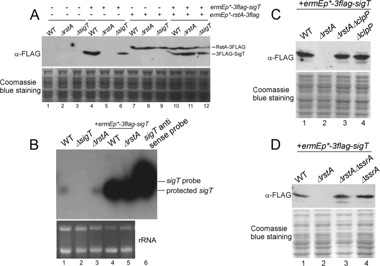
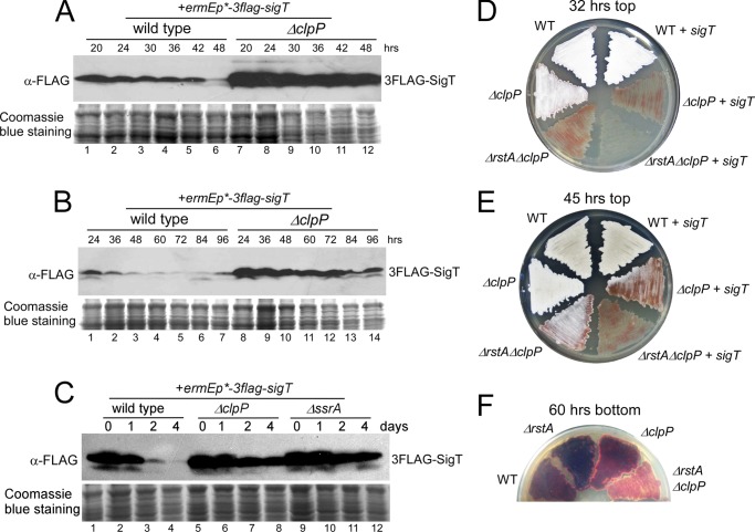
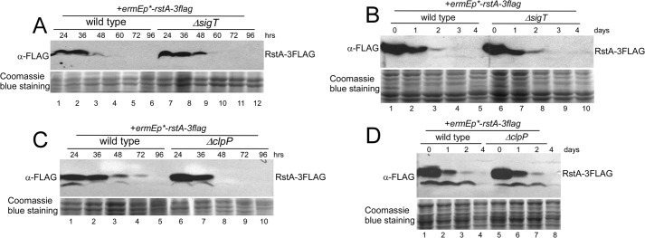
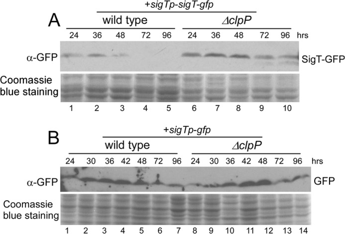
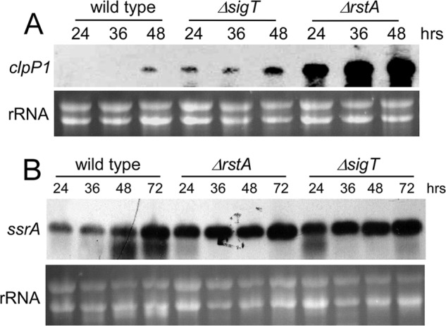
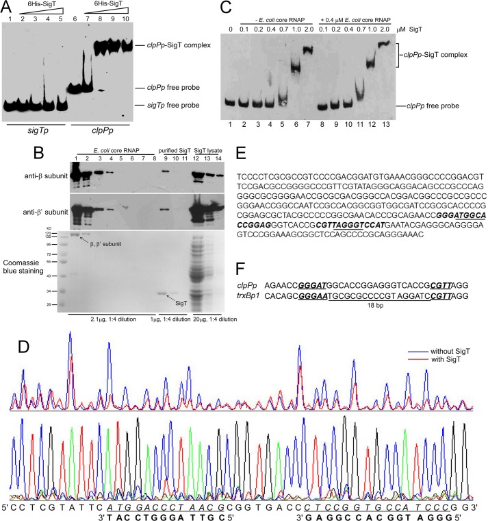
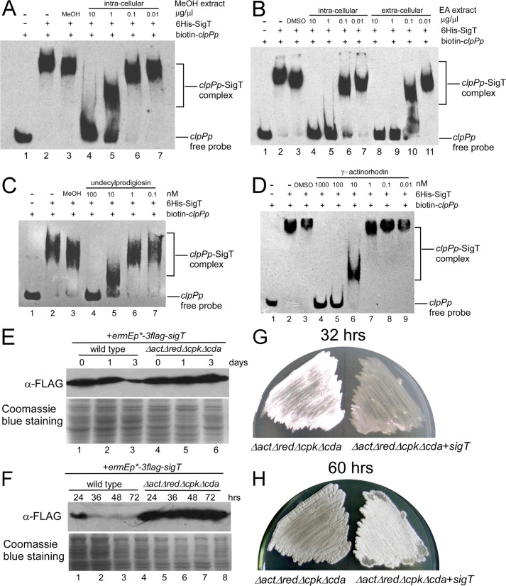
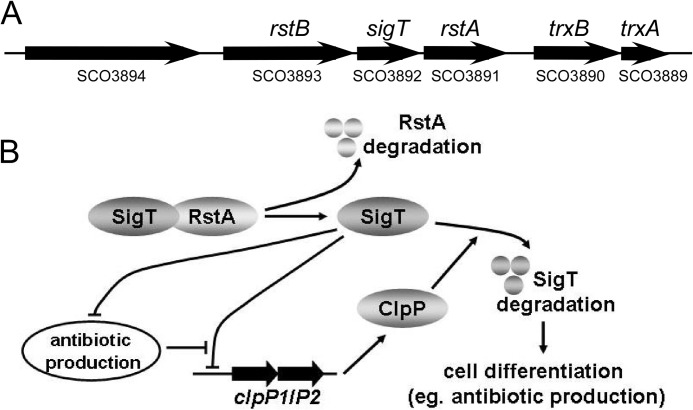
Here we report that in Streptomyces coelicolor, the protein stability of an ECF σ factor SigT, which is involved in the negative regulation of cell differentiation, was completely dependent on its cognate anti-σ factor RstA. The degradation of RstA caused a ClpP/SsrA-dependent degradation of SigT during cell differentiation. This was consistent with the delayed morphological development or secondary metabolism in the ΔclpP background after rstA deletion or sigT overexpression. Meanwhile, SigT negatively regulated clpP/ssrA expression by directly binding to the clpP promoter (clpPp). The SigT-clpPp interaction could be disrupted by secondary metabolites, giving rise to the stabilized SigT protein and retarded morphological development in a non-antibiotic-producing mutant. Thus a novel regulatory mechanism was revealed that the protein degradation of the ECF σ factor was initiated by the degradation of its anti-σ factor, and was accelerated in a dual positive feedback manner, through regulation by secondary metabolites, to promote rapid and irreversible development of the secondary metabolism. This ingenious cooperation of intracellular components can ensure economical and exquisite control of the ECF σ factor protein level for the proper cell differentiation in Streptomyces.

Keywords: Actinobacteria; Antibiotics; Protein Degradation; Secondary Metabolism; Transcription Regulation.


Figures

FIGURE 1.
FIGURE 1.
ClpP/SsrA is involved in degradation of SigT without RstA. A, complementation of RstA for SigT protein stability. Total protein was prepared from strains M145 (wild type, WT), LM22 (ΔrstA), LM21 (ΔsigT) containing either plasmid pL86 (3flag-sigT) or plasmid pL148 (rstA-3flag) or both, in TSB liquid medium for 2 days. SigT and RstA proteins were analyzed by Western blot with α-FLAG antibody. Coomassie Blue staining of total protein served as a loading control. B, sigT expression profile in wild type M145 (WT), LM21 (ΔsigT), LM22 (ΔrstA), M145/pL86 (WT + 3flag-sigT), and LM22/pL86 (ΔrstA + 3flag-sigT) as demonstrated by a ribonuclease protection assay. rRNA was shown as a loading control in a formaldehyde gel. C and D, ClpP/SsrA is required for SigT degradation without RstA. Total protein was prepared from strains M145/pL86 (wild type + 3flag-sigT), LM22/pL86 (ΔrstA + 3flag-sigT), L43/pL86 (ΔrstAΔclpP + 3flag-sigT), and L45/pL86 (ΔclpP + 3flag-sigT) (C), strains M145/pL86 (wild type + 3flag-sigT), LM22/pL86 (ΔrstA + 3flag-sigT), L44/pL86 (ΔrstAΔssrA + 3flag-sigT), and L46/pL86 (ΔssrA + 3flag-sigT) (D) in TSB liquid medium for 2 days and analyzed as in A.
FIGURE 2.
FIGURE 2.
SigT is degraded dependent on ClpP/SsrA during cell differentiation. A–C, ClpP protease is required for SigT degradation during cell differentiation. SigT protein dynamics of strains M145/pL86 (wild type + 3flag-sigT) and L45/pL86 (ΔclpP + 3flag-sigT) on R2YE medium covered with cellophane (A and B) or in a liquid R5-medium (C) for the times indicated as demonstrated by Western blot. Coomassie Blue staining of total protein served as a loading control. Spores of strains M145 (wild type, WT), M145/pL86 (WT + sigT) L45 (ΔclpP), L43 (ΔrstAΔclpP), L45/pL86 (ΔclpP + sigT), and L43/pL86 (ΔrstAΔclpP + sigT) were streaked on R2YE medium for 32 (D) or 45 h (E), respectively, and photographed. F, spores of strains M145 (wild type, WT), LM22 (ΔrstA), L45 (ΔclpP), and L43 (ΔrstAΔclpP) were streaked on R2YE medium for 60 h and photographed.
FIGURE 3.
FIGURE 3.
A and B, RstA degradation independent of SigT during cell differentiation. RstA protein dynamics in strains M145/pLM36 (wild type + rstA-3flag) and LM21/pLM36 (ΔsigT + rstA-3flag) on R2YE medium (A) or in liquid R5-medium (B) for the times indicated as demonstrated by Western blot with α-FLAG antibody. C and D, RstA degradation independent of ClpP protease. RstA protein dynamics in strains M145/pL36 (wild type + rstA-3flag) and L45/pL36 (ΔclpP + rstA-3flag) on R2YE medium (C) or in liquid R5-medium (D) for the times indicated as demonstrated by Western blot. Coomassie Blue staining of total protein served as a loading control.
FIGURE 4.
FIGURE 4.
Endogenous SigT protein dynamics. Protein dynamics of SigT-GFP fusion protein (A) and GFP expressed under sigTp (B) in strains M145/pL97 (wild type + sigTp-sigT-gfp) and L45/pL97 (ΔclpP + sigTp-sigT-gfp) (A), in strains M145/pL96 (wild type + sigTp-gfp) and L45/pL96 (ΔclpP + sigTp-gfp) (B). Cells were collected on R2YE medium overlaid with cellophane after the indicated times and lysed for Western blot with α-GFP antibody. Coomassie Blue staining of total protein was used as a loading control.
FIGURE 5.
FIGURE 5.
A and B, SigT negatively regulates clpP1/P2 and ssrA expression. Ribonuclease protection assay of clpP1 expression (A) and Northern blot analysis of ssrA expression (B) in M145 (wild type), LM21 (ΔsigT) and LM22 (ΔrstA) grown on R2YE medium overlaid with cellophane at various time points. About 10 μg of total RNA run in a formaldehyde gel was for a loading control.
FIGURE 6.
FIGURE 6.
SigT recognizes clpP promoter. A, EMSA for SigT-clpPp binding. About 1 ng of 5′-biotin-labeled sigTp and clpPp probes were incubated with an increasing amount of purified SigT protein, subjected to electrophoresis, and detected by ECL. B, evaluation of RNAP in purified SigT. About 2.1 μg of purified E. coli RNAP core enzyme (lanes 1-8), 1.0 μg of purified SigT (lanes 9-11), and 20 μg of E. coli lysate containing SigT (lanes 12-14) were loaded on a 6% SDS-PAGE for Western blot with antibodies against β and β′ subunits of E. coli RNAP, respectively, or on 12% SDS-PAGE for Coomassie Blue staining. All samples were diluted by 1:4. C, E. coli RNAP core enzyme was not required for SigT-clpPp binding. EMSA of SigT-clpPp binding with an increased amount of purified SigT as indicated without core enzyme (lanes 1-7) or with core enzyme (lanes 8-14). D, DNase I footprinting assay to determine the SigT recognition site on clpPp. 5′-FAM-labeled clpPp probe amplified from pL87 was used in the DNase I footprinting assay with purified SigT. The bottom strand of clpPp was analyzed. The protected region is underlined and in italics. The complementary strand is also shown. The sequencing ladder was generated with the same 5′-FAM-labeled primer. E, the promoter region of clpPp. The SigT binding site deduced from the DNase I footprinting assay is in italics and bold, and the −10 and −35 regions are underlined. F, alignment of recognition sites of SigT on clpPp with trxBp1. The conserved element is bold italic and underlined. The 18-bp interval region between two recognition sites is also shown.
FIGURE 7.
FIGURE 7.
A–D, inhibition of SigT-clpPp binding by secondary metabolites. Intracellular crude extracts with methanol (MeOH) from wild type cells (A), intracellular or extracellular crude extracts with ethyl acetate (EA) from wild type cells (B), HPLC-purified undecylprodigiosin (C) or γ-actinorhodin (D) was incubated with SigT in a concentration gradient before addition of biotin-labeled clpPp probe, respectively, and subjected to EMSA. Methanol or DMSO were used as the solution control. E–H, SigT is stabilized in non-antibiotic-producing cells. SigT protein dynamics in strains M145/pLM86 (wild type + 3flag-sigT) and M1146/pLM86 (ΔactΔredΔcpkΔcda + 3flag-sigT) grown in liquid R5- (E) or on R2YE (F) medium for the times indicated as demonstrated by Western blot with α-FLAG antibody. Coomassie Blue staining of total protein was for a loading control. Spores of strains LM1146 (ΔactΔredΔcpkΔcda) and M1146/pLM86 (ΔactΔredΔcpkΔcda + sigT) were streaked on R2YE medium for 32 (G) and 60 h (H), respectively, and photographed.
FIGURE 8.
FIGURE 8.
A proposed regulatory model for protein degradation of ECF σ factor SigT during cell differentiation. A, genomic organization of sigT, rstA, and their adjacent genes. B, a proposed model of dual positive feedback regulatory mechanism of SigT degradation dependent on ClpP protease and secondary metabolites.

Similar articles

Cited by

References

    1. Paget M. S., Helmann J. D. (2003) The σ70 family of σ factors. Genome Biol. 4, 203. - PMC - PubMed
    1. Gruber T. M., Gross C. A. (2003) Multiple σ subunits and the partitioning of bacterial transcription space. Annu. Rev. Microbiol. 57, 441–466 - PubMed
    1. Staroń A., Sofia H. J., Dietrich S., Ulrich L. E., Liesegang H., Mascher T. (2009) The third pillar of bacterial signal transduction. Classification of the extracytoplasmic function (ECF) σ factor protein family. Mol. Microbiol. 74, 557–581 - PubMed
    1. Ho T. D., Hastie J. L., Intile P. J., Ellermeier C. D. (2011) The Bacillus subtilis extracytoplasmic function σ factor σ(V) is induced by lysozyme and provides resistance to lysozyme. J. Bacteriol. 193, 6215–6222 - PMC - PubMed
    1. Thackray P. D., Moir A. (2003) SigM, an extracytoplasmic function σ factor of Bacillus subtilis, is activated in response to cell wall antibiotics, ethanol, heat, acid, and superoxide stress. J. Bacteriol. 185, 3491–3498 - PMC - PubMed

Publication types

MeSH terms

LinkOut - more resources