IMD-0354 targets breast cancer stem cells: a novel approach for an adjuvant to chemotherapy to prevent multidrug resistance in a murine model
- PMID: 24014113
- PMCID: PMC3754963
- DOI: 10.1371/journal.pone.0073607
IMD-0354 targets breast cancer stem cells: a novel approach for an adjuvant to chemotherapy to prevent multidrug resistance in a murine model
Abstract
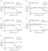
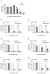
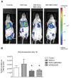
Although early detection of breast cancer improved in recent years, prognosis of patients with late stage breast cancer remains poor, mostly due to development of multidrug resistance (MDR) followed by tumor recurrence. Cancer stem cells (CSCs), with higher drug efflux capability and other stem cell-like properties, are concentrated in a side population (SP) of cells, which were proposed to be responsible for MDR and tumor repopulation that cause patients to succumb to breast cancer. Therefore, targeting of CSCs as an adjuvant to chemotherapy should be able to provide a more effective treatment of this disease. Here, we used IMD-0354, an inhibitor of NF-κB, identified for targeting CSCs, in a combination therapy with doxorubicin encapsulated in targeted nanoparticles. IMD-0354 did target CSCs, evidenced by a decrease in the SP, demonstrated by the inhibition of the following: dye/drug efflux, reduction in ABC transporters as well as in colony formation in soft agar and low attachment plates. Decrease of stem-like gene expression of Oct4, Nanog and Sox2, and apoptosis resistance related to the Survivin gene also was observed after treatment with this compound. In addition, IMD-0354 targeted non-CSCs as indicated by reducing viability and increasing apoptosis. Targeted drug delivery, achieved with a legumain inhibitor, proved to enhance drug delivery under hypoxia, a hallmark of the tumor microenvironment, but not under normoxia. Together, this allowed a safe, non-toxic delivery of both anticancer agents to the tumor microenvironment of mice bearing syngeneic metastatic breast cancer. Targeting both bulk tumor cells with a chemotherapeutic agent and CSCs with IMD-0354 should be able to reduce MDR. This could eventually result in decreasing tumor recurrences and/or improve the outcome of metastatic disease.
Conflict of interest statement
Figures


Similar articles
-
Combination therapy with epigenetic-targeted and chemotherapeutic drugs delivered by nanoparticles to enhance the chemotherapy response and overcome resistance by breast cancer stem cells.J Control Release. 2015 May 10;205:7-14. doi: 10.1016/j.jconrel.2014.11.011. Epub 2014 Nov 15. J Control Release. 2015. PMID: 25445694
-
Indocyanine green/doxorubicin-encapsulated functionalized nanoparticles for effective combination therapy against human MDR breast cancer.Colloids Surf B Biointerfaces. 2019 May 1;177:294-305. doi: 10.1016/j.colsurfb.2019.02.001. Epub 2019 Feb 5. Colloids Surf B Biointerfaces. 2019. PMID: 30771581
-
Histone deacetylase (HDAC) inhibitors and doxorubicin combinations target both breast cancer stem cells and non-stem breast cancer cells simultaneously.Breast Cancer Res Treat. 2020 Feb;179(3):615-629. doi: 10.1007/s10549-019-05504-5. Epub 2019 Nov 29. Breast Cancer Res Treat. 2020. PMID: 31784862
-
Tumor microenvironment and epithelial mesenchymal transition as targets to overcome tumor multidrug resistance.Drug Resist Updat. 2020 Dec;53:100715. doi: 10.1016/j.drup.2020.100715. Epub 2020 Jun 20. Drug Resist Updat. 2020. PMID: 32679188 Review.
-
Multidrug-resistant cancer cells and cancer stem cells hijack cellular systems to circumvent systemic therapies, can natural products reverse this?Cell Mol Life Sci. 2017 Mar;74(5):777-801. doi: 10.1007/s00018-016-2362-3. Epub 2016 Sep 12. Cell Mol Life Sci. 2017. PMID: 27622244 Free PMC article. Review.
Cited by
-
Targeting the NFκB signaling pathways for breast cancer prevention and therapy.Curr Med Chem. 2015;22(2):264-89. doi: 10.2174/0929867321666141106124315. Curr Med Chem. 2015. PMID: 25386819 Free PMC article. Review.
-
Targeting STING pathways for the treatment of cancer.Oncoimmunology. 2015 Jan 7;4(12):e988463. doi: 10.4161/2162402X.2014.988463. eCollection 2015 Dec. Oncoimmunology. 2015. PMID: 26587334 Free PMC article.
-
STING ligand c-di-GMP improves cancer vaccination against metastatic breast cancer.Cancer Immunol Res. 2014 Sep;2(9):901-10. doi: 10.1158/2326-6066.CIR-13-0123. Epub 2014 Jun 9. Cancer Immunol Res. 2014. PMID: 24913717 Free PMC article.
-
β-arrestin 2 is associated with multidrug resistance in breast cancer cells through regulating MDR1 gene expression.Int J Clin Exp Pathol. 2015 Feb 1;8(2):1354-63. eCollection 2015. Int J Clin Exp Pathol. 2015. PMID: 25973019 Free PMC article.
-
Exosome-mediated peritoneal dissemination in gastric cancer and its clinical applications.Biomed Rep. 2018 Jun;8(6):503-509. doi: 10.3892/br.2018.1088. Epub 2018 Apr 19. Biomed Rep. 2018. PMID: 29774141 Free PMC article. Review.
References
-
- Siegel R, Naishadham D, Jemal A (2012) Cancer statistics, 2012. CA Cancer J Clin 62: 10-29. doi:10.3322/caac.20138. PubMed: 22237781. - DOI - PubMed
-
- Octavia Y, Tocchetti CG, Gabrielson KL, Janssens S, Crijns HJ et al. (2012) Doxorubicin-induced cardiomyopathy: from molecular mechanisms to therapeutic strategies. J Mol Cell Cardiol 52: 1213-1225. doi:10.1016/j.yjmcc.2012.03.006. PubMed: 22465037. - DOI - PubMed
-
- Ailles LE, Weissman IL (2007) Cancer stem cells in solid tumors. Curr Opin Biotechnol 18: 460-466. doi:10.1016/j.copbio.2007.10.007. PubMed: 18023337. - DOI - PubMed
-
- Dean M (2009) ABC transporters, drug resistance, and cancer stem cells. J Mammary Gland Biol Neoplasia 14: 3-9. doi:10.1007/s10911-009-9109-9. PubMed: 19224345. - DOI - PubMed
Publication types
MeSH terms
Substances
Grants and funding
LinkOut - more resources
Full Text Sources
Other Literature Sources
Research Materials

