

Case Reports
doi: 10.1136/bcr-2013-200268.
Osteoma of temporomandibular joint: a rarity
Affiliations
- PMID: 24014334
- PMCID: PMC3794170
- DOI: 10.1136/bcr-2013-200268
Item in Clipboard
Case Reports
Osteoma of temporomandibular joint: a rarity
BMJ Case Rep.
.
Abstract
Osteoma is a benign tumour consisting of mature bone tissue. It is an uncommon lesion that occurs in the bones of the craniofacial complex. Only a few cases involving the temporomandibular joint have been reported. An osteoma of the left temporomandibular joint causing limited mouth opening in a 22-year-old man with CT findings revealing the unusual possibility in differential diagnosis of trismus.
Figures
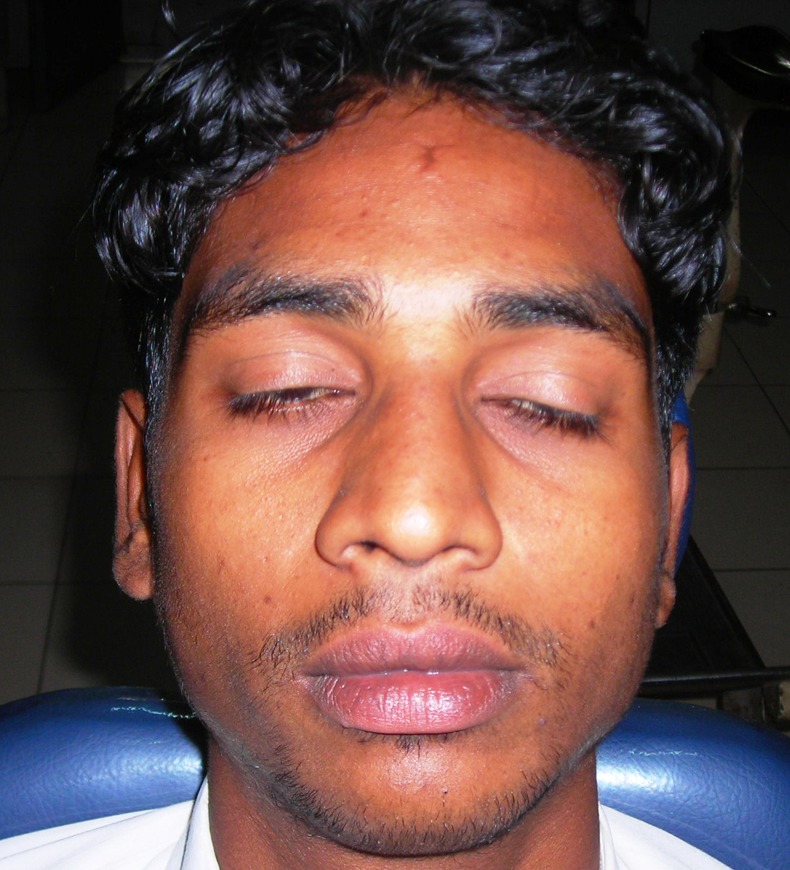
Extra oral picture of patient.
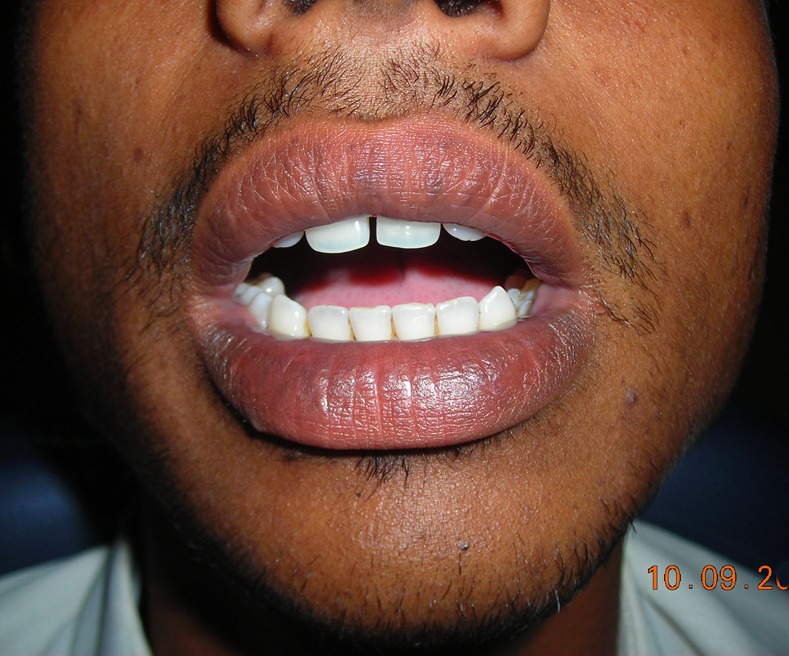
Restricted mouth opening.
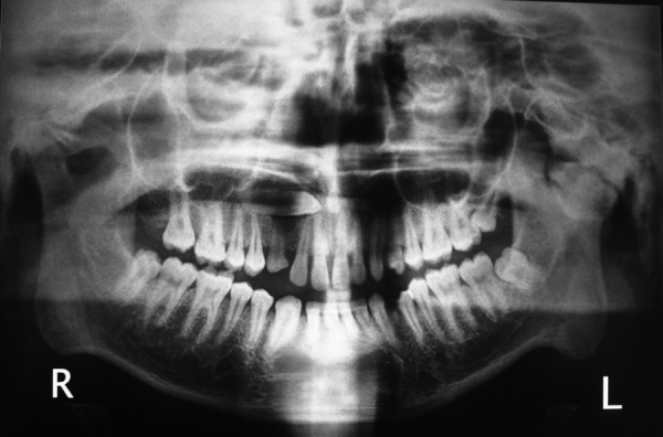
Orthopantomograph showing large radiopacity in the left temporomandibular joint region.
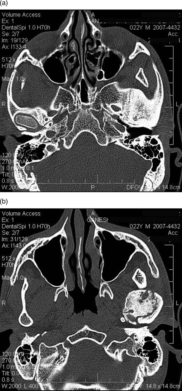
(A) Axial CT slices showing hyperdense mass of size 2.84 cm mediolaterally in left condylar region involving the sphenoid and temporal bones and (B) multiple hypodense areas, within the mass.
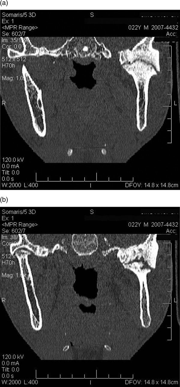
(A) Frontal slices showing hyperdense enlarged temporomandibular joint components with linear hypodense area of variable thickness and (B) hypodense area of variable thickness joint space.
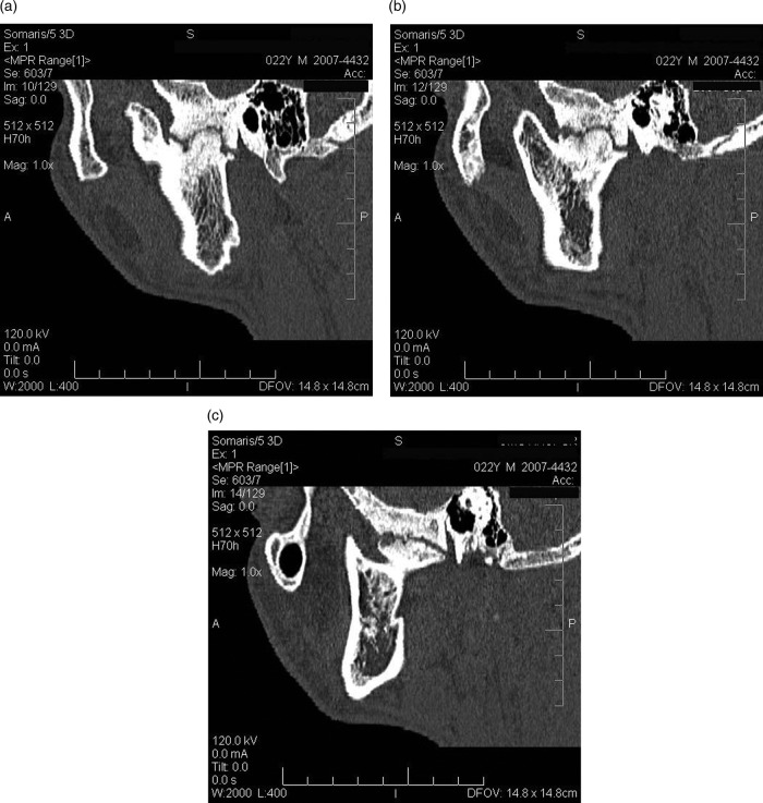
(A) Sagittal slices showing obliterated joint space (B) change in the structure of the temporomandibular joint components and (C) increased size of the left coronoid process.
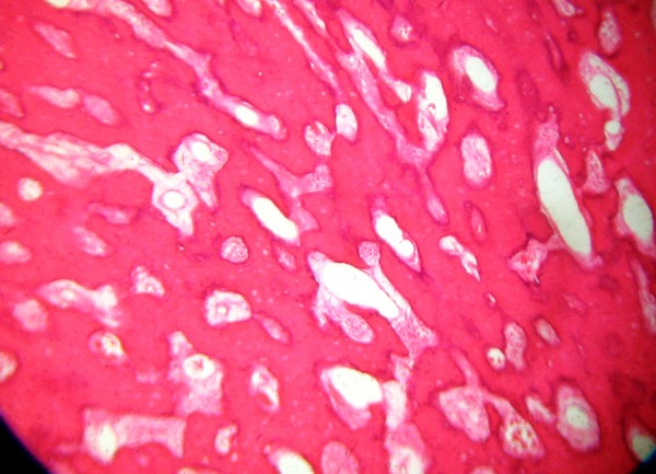
H&E-stained section showing mature lamellated compact bone.
References
-
- Park W, Nam W, Park H, et al. Intraosseous lesion in mandibular condyle mimicking temporomandibular disorders: report of 3 cases. J Orofac Pain 2008;2013:65–70 - PubMed
-
- Siar CH, Jalil AA, Ram S, et al. Osteoma of the condyle as the cause of limited mouth opening: a case report. J Oral Sci 2004;2013:51–3 - PubMed
-
- Yonezu H, Wakoh M, Otonari T, et al. Osteoma of mandibular condyle as cause of acute pain & limited mouth opening: case report. Bull Tokyo Dent Coll 2007;2013:193–7 - PubMed
-
- Yang C, Qiu WL. Osteoid osteoma of the eminence of temporomandibular joint. Br J Oral Maxillofac Surg 2001;2013:404–6 - PubMed
-
- Kademani D, Bevin C. A mass in the temporomandibular joint. J Am Dent Assoc 2008;2013:301–3 - PubMed
Publication types
MeSH terms
LinkOut - more resources
Full Text Sources
Other Literature Sources
Medical