Review
. 2013 Jul;2(4):427-42.
doi: 10.1002/wdev.97. Epub 2012 Nov 26.

Birth defects associated with perturbations in preimplantation, gastrulation, and axis extension: from conjoined twinning to caudal dysgenesis

Affiliations
Review

Birth defects associated with perturbations in preimplantation, gastrulation, and axis extension: from conjoined twinning to caudal dysgenesis

Anna Ferrer-Vaquer et al. Wiley Interdiscip Rev Dev Biol. 2013 Jul.

Abstract

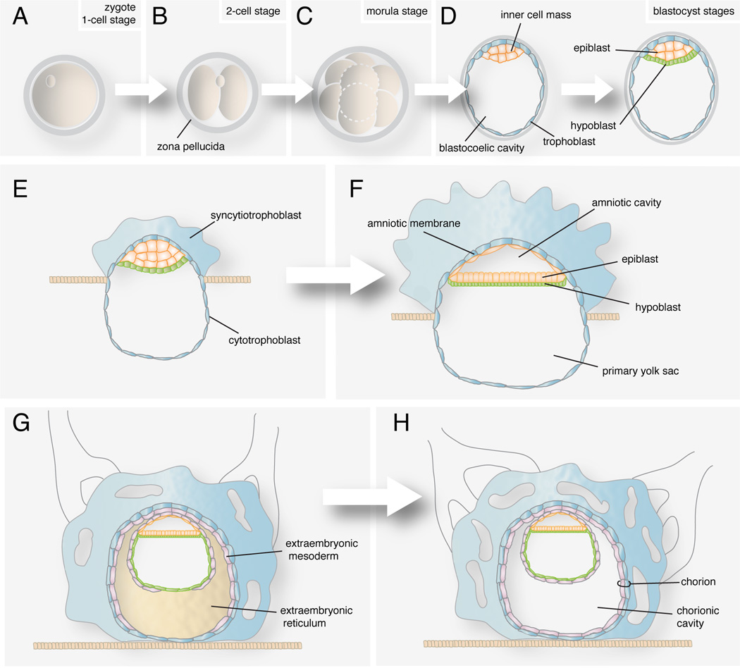
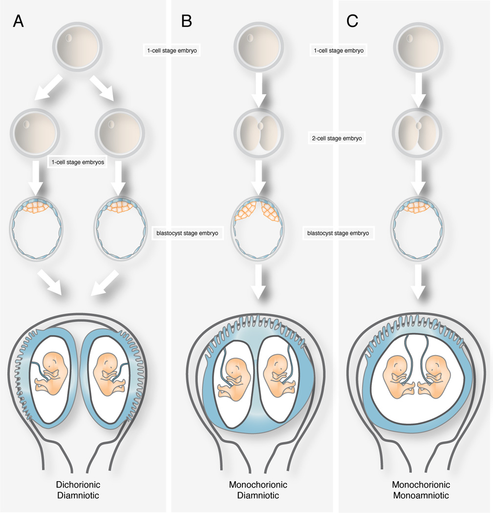
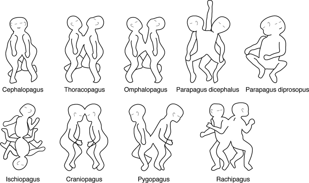
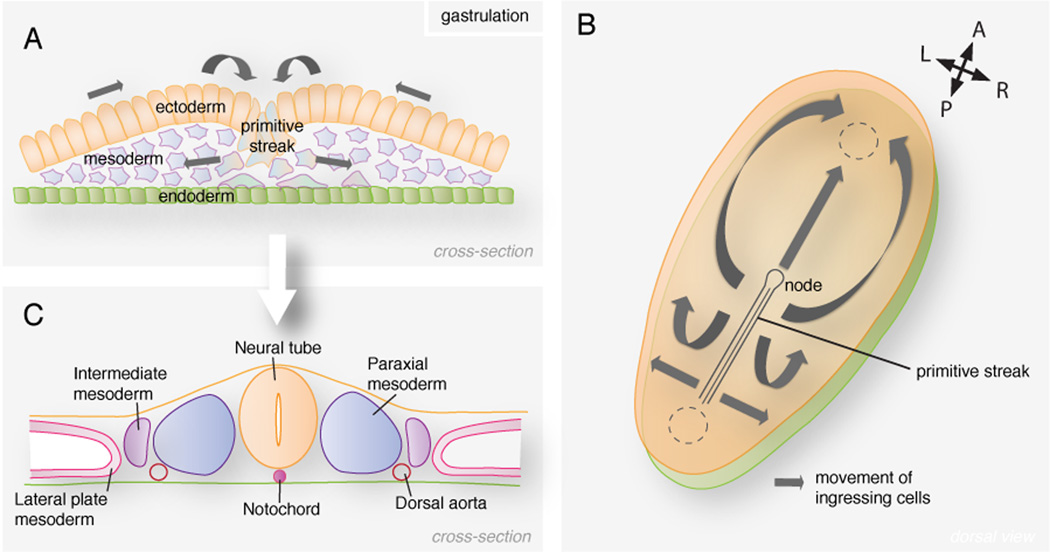
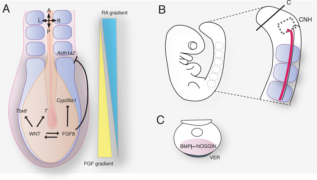
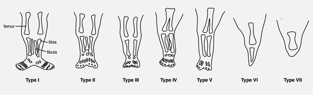
Congenital malformations represent approximately 3 in 100 live births within the human population. Understanding their pathogenesis and ultimately formulating effective treatments are underpinned by knowledge of the events and factors that regulate normal embryonic development. Studies in model organisms, primarily in the mouse, the most prominent genetically tractable mammalian model, have equipped us with a rudimentary understanding of mammalian development from early lineage commitment to morphogenetic processes. In this way, information provided by studies in the mouse can, in some cases, be used to draw parallels with other mammals, including human. Here, we provide an overview of our current understanding of the general sequence of developmental events from early cell cleavages to gastrulation and axis extension occurring in human embryos. We will also review some of the rare birth defects occurring at these stages, in particular those resulting in conjoined twinning or caudal dysgenesis.


Figures

FIGURE 1
FIGURE 1. Events occurring the first two weeks of human development
(A) Zygote. (B) Two-cell stage embryo. (C) Morula before compaction. (D) Blastocyst forms after compaction and formation of the blastocoel. The embryo segregates into epiblast and hypoblast. (E) Implantation starts at the end of the first week. The syncytiotrophoblast begins to invade the uterine wall. (F) The amniotic cavity appears. (G) Hypoblast cells invade the lining of the primary yolk sac. Extraembryonic reticulum forms between the hypoblast and cytotrophoblast. Extraembryonic mesoderm colonizes the reticulum. (H) The reticulum cavitates leaving the chorionic cavity. The extraembryonic mesoderm and underlying cytotrophoblast form the chorion.
FIGURE 2
FIGURE 2. Uterine disposition of monozygotic twins
(A) Dichorionic diamniotic twins develop from a single zygote divided before the inner cell mass forms. (B) Monochorionic diamniotic twins form when the inner cell mass divides between the third and eighth day of development. (C) Monochorionic monoamniotic twins develop from the division of the bilaminar disc.
FIGURE 3
FIGURE 3. Classification of conjoined twins according to Spencer
FIGURE 4
FIGURE 4. Mesoderm formation in the mammalian embryo
(A) Cells in the epiblast undergo an epithelial to mesenchymal transition at the primitive streak and colonize the space between the epiblast and hypoblast. (B) Ingressing cells migrate in all directions. Cells ingressing through the node migrate anteriorly forming the notochord. Cells ingressing through the streak and migrating cranially contribute to the head and cardiac mesoderm. Epiblast cells migrating laterally form the mesoderm at each side of the midline. (C) The different mesodermal lineages are established along the mediolateral axis of the embryo.
FIGURE 5
FIGURE 5. Signals that regulate axial elongation in the mouse embryo
(A) Molecular pathways acting at the posterior growth region and contributing to the antagonistic gradients of FGF and RA. (B) Mouse embryo at embryonic day 9. The chordoneural hinge (CNH) is located caudally and lies contiguous with the notochord and neural tube. (C) Noggin expression in the ventral ectodermal ridge (VER) controls the level of BMP signaling required for caudal ventral mesoderm morphogenesis.
FIGURE 6
FIGURE 6. Classification of sirenomelia by the presence or absence of bones in the lower limb according to Stocker and Heifetz
Adapted from with permission.

Similar articles

Cited by

References

    1. Thomson JA, Itskovitz-Eldor J, Shapiro SS, Waknitz MA, Swiergiel JJ, Marshall VS, Jones JM. Embryonic stem cell lines derived from human blastocysts. Science. 1998;282:1145–1147. - PubMed
    1. Yu J, Thomson JA. Pluripotent stem cell lines. Genes Dev. 2008;22:1987–1997. - PMC - PubMed
    1. Rossant J, Tam PP. Blastocyst lineage formation, early embryonic asymmetries and axis patterning in the mouse. Development. 2009;136:701–713. - PubMed
    1. Cockburn K, Rossant J. Making the blastocyst: lessons from the mouse. J Clin Invest. 2010;120:995–1003. - PMC - PubMed
    1. Lanner F, Rossant J. The role of FGF/Erk signaling in pluripotent cells. Development. 2010;137:3351–3360. - PubMed

Publication types

Supplementary concepts

LinkOut - more resources