Review
. 2013 Jul;2(4):443-59.
doi: 10.1002/wdev.88. Epub 2012 Sep 18.

Neocortical neurogenesis and neuronal migration

Affiliations
Review

Neocortical neurogenesis and neuronal migration

Xin Tan et al. Wiley Interdiscip Rev Dev Biol. 2013 Jul.

Abstract

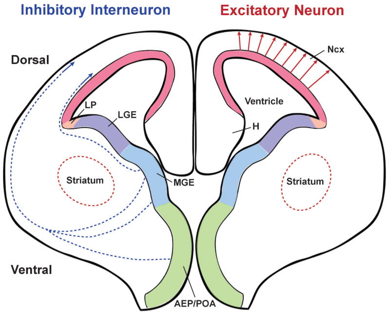
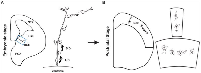
The neocortex, the evolutionarily newest part of the cerebral cortex, controls nearly all aspects of behavior, including perception, language, and decision making. It contains an immense number of neurons that can be broadly divided into two groups, excitatory neurons and inhibitory interneurons. These neurons are predominantly produced through extensive progenitor cell divisions during the embryonic stages. Moreover, they are not randomly dispersed, but spatially organized into horizontal layers that are essential for neocortex function. The formation of this laminar structure requires exquisite control of neuronal migration from their birthplace to their final destination. Extensive research over the past decade has greatly advanced our understanding of the production and migration of both excitatory neurons and inhibitory interneurons in the developing neocortex. In this review, we aim to give an overview on the molecular and cellular processes of neocortical neurogenesis and neuronal migration.


Figures

Figure 1
Figure 1. Distinct origins of excitatory neurons and inhibitory interneurons in the developing mouse neocortex
Excitatory neurons are generated in the proliferative zone of the dorsal telencephalon and then migrate radially to the cortical plate (red arrows). In contrast, inhibitory interneurons are produced in the proliferative zone of the ventral telencephalon, especially the MGE and AEP/POA, and migrate tangentially to reach the neocortex following two major routes (blue dashed arrows). The colored regions indicate the proliferative zones across the embyronic brain expressing different transcription factors that are essential for proper neurogenesis of distinct neuronal populations. AEP: anterior entopeduncular area; H: hippocampus; LGE: lateral ganglionic eminence; LP: lateral pallium; MGE: medial ganglionic eminence; Ncx: neocortex; POA: preoptic area.
Figure 2
Figure 2. Diverse populations of excitatory neuron progenitor cells in the mouse neocortex
During early brain development, neuroepithelial cells (NECs) are the major neural progenitors in the neocortex. NECs divide symmetrically to generate additional NECs, some of which give rise to the first group of neurons (N) through asymmetric division. As the developing brain epithelium thickens, NECs elongate and transit to radial glial cells (RGCs). RGCs can also divide symmetrically to expand the progenitor pool or asymmetrically to generate neurons either directly or indirectly through intermediate progenitor cells (IPCs), which generate neurons directly through symmetric division in the subventricular zone (SVZ). It remains unclear how short neural progenitors (SNPs) are generated. It is possible that they are generated from RGCs or they may be a distinct population originated directly from NECs. Most SNPs produce postmitotic neurons directly in the ventricular zone (VZ). Outer subventricular zone (OSVZ) progenitors likely originate from RGCs through oblique division, which leads to loss of the apical process and ascension of the nucleus towards the cortical plate/intermediate zone (CP/IZ). The minor population of OSVZ progenitors can also generate neurons through asymmetric division outside of the VZ.
Figure 3
Figure 3. Models of symmetric and asymmetric divisions of radial glial progenitor cells in the mouse neocortex
(A) Cleavage-plane orientation model. When the cleavage-plane is perpendicular to the VZ surface, the division of RGCs results in symmetric inheritance of critical fate determinants enriched at the VZ surface (represented by a green line) and consequently the same daughter cell fate specification (i.e. symmetric division). When the cleavage-plane is parallel to the VZ surface, the division results in asymmetric inheritance of critical fate determinants enriched at the VZ surface and consequently distinct daughter cell fate specification - one remains as RGC whereas the other becomes a postmitotic neuron (N) or an intermediate progenitor (I) (i.e. asymmetric division). (B) Oblique division model. Dividing progenitor cells possess a small patch of membrane close to the VZ surface (represented by a short purple line), the inheritance of which is critical for fate specification. Bisection of this small membrane patch leads to symmetric division, whereas a slight tilt in the vertical cleavage plane (i.e. oblique division) bypasses it and results in asymmetric division. (C) Dynamic polarity establishment model. The distribution of the evolutionarily conserved cell polarity protein PARD3 is dynamic depending on cell cycle progression, which becomes dispersed during mitosis. Even distribution of PARD3 in dividing RGCs leads to symmetric division, whereas polarized distribution of PARD3 results in its asymmetric inheritance by the two daughter cells, followed by differential activation of NOTCH signaling and subsequent distinct fate specification. (D) Basal process inheritance model. Inheritance of the basal process has been linked to fate specification though it is still controversial. The basal process can be bisected and inherited by both daughter cells during the symmetric division, although it has also been postulated that the basal process may be inherited by only one daughter RGC while the other has to elaborate a new basal process. For the asymmetric division, earlier studies suggested that the daughter cell that inherits the basal process becomes a differentiating neuron whereas the other daughter grows a new process and remains in the VZ as a progenitor. Recent work indicated that the daughter cell inheriting the basal process remains a progenitor (either a RGC or OSVZ progenitor) while the other differentiates (either a neuron or an intermediate progenitor).
Figure 4
Figure 4. Inside-out layer formation of excitatory neurons in the neocortex
The PP is formed by the production of the first wave of post-mitotic neurons that migrate from the VZ to the pial surface. Then, a second wave of new-born neurons (blue triangles) migrate through the SVZ/IZ and split the PP into the more superficial MZ (neurons in the MZ are represented by orange ovals) and the more deeply located SP (neurons in the SP are represented by green diamonds), creating the CP – the future neocortex. Neurons generated subsequently (represented by triangles with different colors, pink, purple and yellow by birth order) expand the CP in an inside-out fashion, as later-born neurons pass the existing neurons to occupy more superficial layers. The VZ progressively shrinks as the neural progenitor cells decrease during later embryonic development. During the postnatal development towards adulthood, the SP degenerates and leaves behind a six-layered neocortex (panel on the right). CP: cortical plate; IZ: intermediate zone; MZ: marginal zone; PP: preplate; SP: subplate; SVZ: subventricular zone; VZ: ventricular zone; WM: white matter.
Figure 5
Figure 5. Clonal production and organization of inhibitory interneurons in the mouse neocortex
(A) At embryonic stage, RGCs in the VZ of the MGE divide asymmetrically to self-renew and to simultaneously produce differentiating interneurons or IPCs that divide symmetrically in the SVZ to produce differentiating interneurons. The progeny of the same RGC initially migrate along the mother RGC and form radially aligned clonal clusters (the clone in the MGE is shown at higher magnification in the right panel). The new-born cells progressively move away from the VZ, differentiate, and migrate tangentially towards the neocortex. (B) After arriving at their destination in the neocortex, inhibitory interneuron clones do not randomly disperse, but form spatially organized clusters which are either vertically or horizontally oriented (one example in each case is shown at higher magnification in the right panel). A.D., asymmetric division; S.D., symmetric division. Adapted from Fig. S14 .

Similar articles

Cited by

References

    1. Gotz M, Huttner WB. The cell biology of neurogenesis. Nat Rev Mol Cell Biol. 2005;6:777–788. - PubMed
    1. Williams BP, Price J. Evidence for multiple precursor cell types in the embryonic rat cerebral cortex. Neuron. 1995;14:1181–1188. - PubMed
    1. Hartfuss E, Galli R, Heins N, Gotz M. Characterization of CNS precursor subtypes and radial glia. Dev Biol. 2001;229:15–30. - PubMed
    1. Malatesta P, Hack MA, Hartfuss E, Kettenmann H, Klinkert W, Kirchhoff F, Gotz M. Neuronal or glial progeny: regional differences in radial glia fate. Neuron. 2003;37:751–764. - PubMed
    1. Aaku-Saraste E, Hellwig A, Huttner WB. Loss of occludin and functional tight junctions, but not ZO-1, during neural tube closure--remodeling of the neuroepithelium prior to neurogenesis. Dev Biol. 1996;180:664–679. - PubMed

Publication types

LinkOut - more resources