Review
. 2013 Jul;2(4):461-78.
doi: 10.1002/wdev.89. Epub 2012 Oct 4.

Genetic causes of microcephaly and lessons for neuronal development

Affiliations
Review

Genetic causes of microcephaly and lessons for neuronal development

Edward C Gilmore et al. Wiley Interdiscip Rev Dev Biol. 2013 Jul.

Abstract

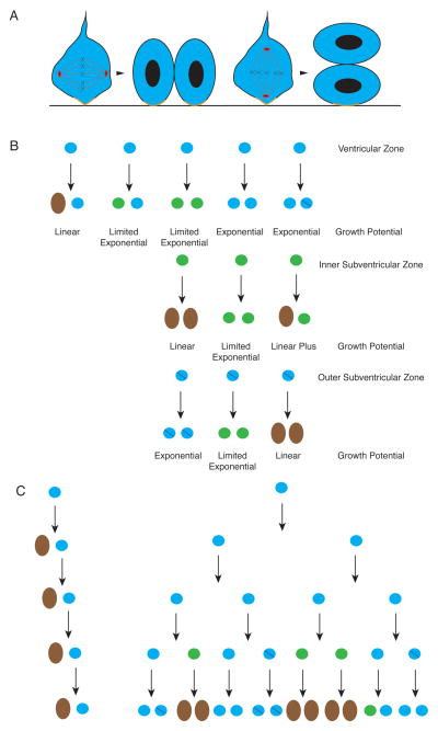
The study of human developmental microcephaly is providing important insights into brain development. It has become clear that developmental microcephalies are associated with abnormalities in cellular production, and that the pathophysiology of microcephaly provides remarkable insights into how the brain generates the proper number of neurons that determine brain size. Most of the genetic causes of 'primary' developmental microcephaly (i.e., not associated with other syndromic features) are associated with centrosomal abnormalities. In addition to other functions, centrosomal proteins control the mitotic spindle, which is essential for normal cell proliferation during mitosis. However, the brain is often uniquely affected when microcephaly genes are mutated implying special centrosomal-related functions in neuronal production. Although models explaining how this could occur have some compelling data, they are not without controversy. Interestingly, some of the microcephaly genes show evidence that they were targets of evolutionary selection in primates and human ancestors, suggesting potential evolutionary roles in controlling neuronal number and brain volume across species. Mutations in DNA repair pathway genes also lead to microcephaly. Double-stranded DNA breaks appear to be a prominent type of damage that needs to be repaired during brain development, yet why defects in DNA repair affect the brain preferentially and if DNA repair relates to centrosome function, are not clearly understood.


Figures

Figure 1
Figure 1
MRI images from two 12 month old children, the top with developmental microcephaly and bottom with a head circumference in normal range. The images are T1 weighted sequence in mid-sagittal and axial planes. Note the dramatic reduction in brain volume with the relative preservation in structure and size of facial features. The images are scaled to the same size (bar is 5 cm).
Figure 2
Figure 2
(A) Examples of head circumference growth curves for Boys aged 0 (Birth) to 18 years (in months). The mean with 2 standard deviations above (+2 SD) and below (−2 SD) the mean to illustrate normal growth patterns. Note the very rapid expansion in head circumference during the first year of life. The purple line with Xs shows, a child with a developmental microcephaly that starts below the normal growth curves. The head growth remain below the normal curves and follow it’s own trajectory with a shallower slope due to diminished brain growth potential. Clinically, these children often gain milestones more slowly compared to other children and the developmental potential eventually plateaus. However, there is no developmental milestone loss unless there are additional complications, neurological or otherwise. An example of a child with a degenerative condition leading to microcephaly is shown in light blue with Xs. Note that the child starts within the normal range then starts to cross percentile curves during the mid to late first year of life. Clinically, the child may gain early developmental milestones such as smiling, rolling over and sitting without support but then loses them as the neurodegenerative process proceeded. (B) Pre and post-natal brain growth from ~13 weeks post-fertilization to 36 months after being born at full term (prenatal ages in green and post-natal ages in black). Also shown are qualitative illustrations of the approximate timing of cerebral cortical development of neurons , 172 (yellow), astrocytes (purple) 173, myelination 174 (blue) and synapses175 (green). Developmental processes are demonstrating the relative temporal peak of each process with arbitrary units for the Y-axis while no attempt is made to show the relative contribution to brain volume. The exact extent of each process is not represented as each phenomenon (except for neurogenesis) continues at some level into adolescence. Comparisons between processes is very difficult because were different methods used for each type of study. In addition, some studies measure the density of phenomena in a growing volume further complicating extrapolation. The growth curves are a mathematic composite of fetal 176 and childhood177 data and are intended for illustrative purposes not clinical use.
Figure 3
Figure 3
(A) Illustration of pseudostratified epithelium of the developing cerebral cortex prior to neurogenesis. It has the superficial appearance of layers due to nuclei being scattered from the ventricular to pial surface. However, cells are apparently uniform in character and all retain contacts with both the ventricular and pial surfaces. Within each cell, nuclei move upward toward the pial surface where they synthesize DNA and downward to the pial surface where they divide in mitosis. This pattern of nuclei movement is retained within the ventricular zone later in development described in B. Prior to neurogenesis, nearly all cells are uncommitted progenitors and the proliferative pool exponentially. (B) A diagram of the layering found during neurogenesis to emphasize the proliferative pools. Within the ventricular zone (represented by plain blue circles), uncommitted progenitors divide at the apical surface (dividing cells marked with red dot (indicative of the mitotic marker phospho-histone H3). Some of the progeny will become committed but retain limited proliferative capacity, start to express the marker TBR2, and move the inner subventricular zone, marked in green and also referred to intermediate precursors. They also divide (red dots, phospho-histone H3) but remain in the subventricular zone and do not have a connection with the apical surface. Cells within the outer subventricular zone (blue circles with purple slash) have many of the same markers of the ventricular zone, are uncommitted, have extensive proliferative capacity, but do not have connection with the apical surface, unlike the ventricular zone cells. The intermediate zone is cell sparse and the cortical plate consists of differentiated neurons that will become the cerebral cortex.
Figure 4
Figure 4
A cartoon schematic of centriole biogenesis in dividing cells (see the excellent review for further details). The potential roles of microcephaly related genes are noted. After M phase, in G1 two centrioles (large green boxes) are attached via a linker that contains CDK5RAP2. At the G1-S transition and only once during a cell cycle, a new centriole (small green box) is formed adjacent to parental centriole (larger green box). CEP152 is required for this process and it interacts with CEP63. Centriole duplication potentially involves STIL via its interaction with SAS6. The new centriole remains attached to the older centriole (small orange line). The new centriole elongates during S-phase and CENPJ is potentially involved this activity. MCPH1 may play a role in cell-cycle checkpoint control at the G2/M transition. ASPM may play an important role in mitotic spindle orientation during M phase. The alterations of spindle orientation play an important role in cell fate decisions invertebrates and may play a role in mammalian cortical development. Each cell inherits one centrosome, but the centrioles have different levels of maturity as it takes 1 ½ cell cycles for a centriole to fully mature as it accumulates distal appendages (black lines) and other maturation markers (more mature has asterisks). The differences in maturity between centrioles have potentially important biological consequences.
Figure 5
Figure 5
(A) Illustration of different planes of cell division in the ventricular zone with an emphasis on the retention of the apical membrane complex (orange). When a dividing cell has centrosomes aligned so that the cleavage plane is perpendicular to the ventricular surface, the apical membrane complex is shared equally between progeny. When the centrosomes are aligned so that the cleavage plane is parellel to the ventricular surface, one cell inherits the apical membrane complex while the other does not. Since the apical membrane complex takes up a very small area of the ventricular surface, it has been hypothesized that slight deviations from a perpendicular cleavage plane may be enough to cause unequal distribution in progeny. (B) Illustration of potential progeny derived from different proliferative zones along with the growth potential. For instance, only linear growth occurs when a ventricular zone cell (blue circle) divides to produce one neuron (brown oval) and one uncommitted precursor. However, when two uncommitted precursors (in either ventricular zone (blue circle) or outer subventricular zone (blue circle with purple slash)) cell growth can be exponential. When the progeny include inner subventricular zone cells (green circle), there can be extensive expansion compared to producing neurons directly. However, true exponential growth is not possible because these neuronally committed cells divide a limited number of times before differentiating into neurons. The outer subventricular zone cells appear to be comparable to the proliferative capacity of the ventricular zone, however, it is not fully clear what cells it can produce or how these decisions are controlled. Presumably, fate decisions in this group can significantly alter the final number of neurons in the brain. (C) An example of the number of cells produced in 4 divisions from one ventricular zone cell with linear growth versus combined exponential, limited exponential and linear growth demonstrating how neuronal number could be controlled though control of cell fate. Note, neurons (brown) no longer divide but move out of the ventricular zone and migrate to the cortical plate.
Figure 6
Figure 6
Schematic of DNA damage response to double strand breaks. After a double strand break occurs ATM is activated via interaction with the MRN (MRE11A-RAD50-NBN (NBS1) complex. After ATM is activated it establishes a very broad cascade of second messengers (including the MRN complex) to perform a varieties of cellular functions. Progression of the cell cycle is arrested. This is thought to help prevent the propagation of the DNA damage and allow time for repair. Arrest of the cell cycle involves interaction with the cyclin dependent kinase machinery and potentially the centrosome as well. In addition, apoptotic response pathways related to TP53 (p53) are activated permitting the cell to initiate programmed cell death if the level of DNA damage is severe, but much remains to be determined as to how these decisions are made 178. Finally, the proteins that will perform the DNA repair are attracted to the site of damage break. Abnormalities in genes involved in the non-homologous end-joining pathway leads to microcephaly in humans including LIG4, NHEJ and PNKP and an analogous phenotype in mice Lig4 , , Xrcc4 , Xrcc6 (Ku70)/Xrcc5 (Ku80), or Prkdc (DNA-PKcs) .

References

    1. Dolk H. The predictive value of microcephaly during the first year of life for mental retardation at seven years. Dev Med Child Neurol. 1991;33:974–983. - PubMed
    1. Bilguvar K, Ozturk AK, Louvi A, Kwan KY, Choi M, Tatli B, Yalnizoglu D, Tuysuz B, Caglayan AO, Gokben S, et al. Whole-exome sequencing identifies recessive WDR62 mutations in severe brain malformations. Nature. 2010;467:207–210. - PMC - PubMed
    1. Yu TW, Mochida GH, Tischfield DJ, Sgaier SK, Flores-Sarnat L, Sergi CM, Topcu M, McDonald MT, Barry BJ, Felie JM, et al. Mutations in WDR62, encoding a centrosome-associated protein, cause microcephaly with simplified gyri and abnormal cortical architecture. Nat Genet. 2010;42:1015–1020. - PMC - PubMed
    1. Nicholas AK, Khurshid M, Desir J, Carvalho OP, Cox JJ, Thornton G, Kausar R, Ansar M, Ahmad W, Verloes A, et al. WDR62 is associated with the spindle pole and is mutated in human microcephaly. Nat Genet. 2010;42:1010–1014. - PMC - PubMed
    1. Spalding KL, Bhardwaj RD, Buchholz BA, Druid H, Frisen J. Retrospective birth dating of cells in humans. Cell. 2005;122:133–143. - PubMed

Publication types

LinkOut - more resources