Review
. 2013 Jul;2(4):545-57.
doi: 10.1002/wdev.100. Epub 2012 Nov 16.

Retinal differentiation in Drosophila

Affiliations
Review

Retinal differentiation in Drosophila

Jessica E Treisman. Wiley Interdiscip Rev Dev Biol. 2013 Jul.

Abstract

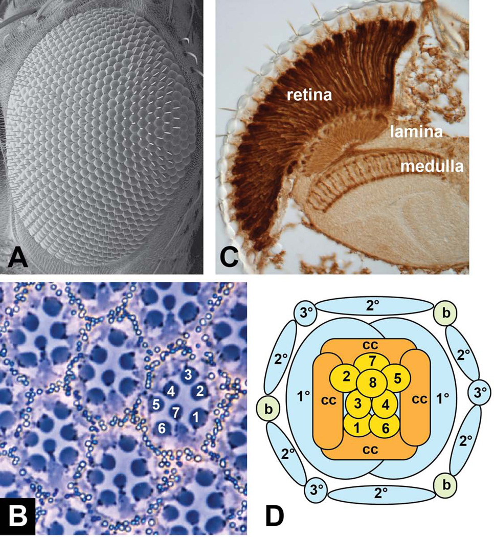
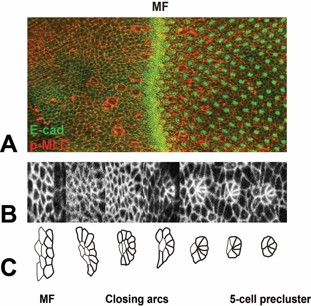
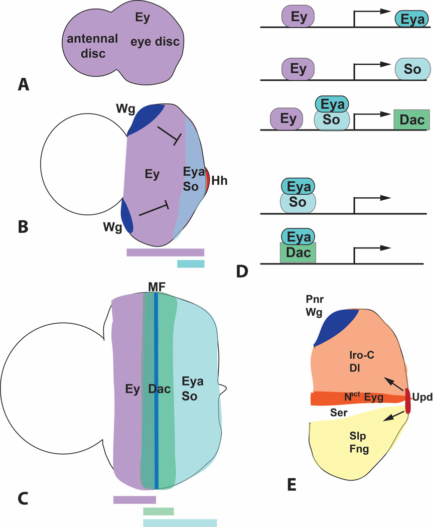
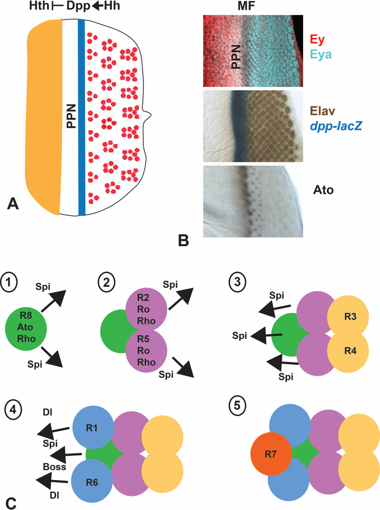
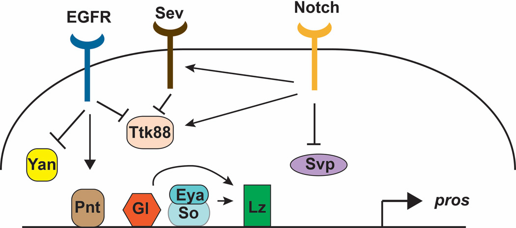
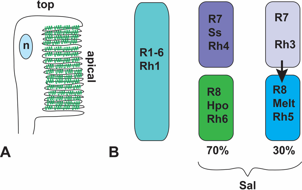
Drosophila eye development has been extensively studied, due to the ease of genetic screens for mutations disrupting this process. The eye imaginal disc is specified during embryonic and larval development by the Pax6 homolog Eyeless and a network of downstream transcription factors. Expression of these factors is regulated by signaling molecules and also indirectly by growth of the eye disc. Differentiation of photoreceptor clusters initiates in the third larval instar at the posterior of the eye disc and progresses anteriorly, driven by the secreted protein Hedgehog. Within each cluster, the combined activities of Hedgehog signaling and Notch-mediated lateral inhibition induce and refine the expression of the transcription factor Atonal, which specifies the founding R8 photoreceptor of each ommatidium. Seven additional photoreceptors, followed by cone and pigment cells, are successively recruited by the signaling molecules Spitz, Delta, and Bride of sevenless. Combinations of these signals and of intrinsic transcription factors give each ommatidial cell its specific identity. During the pupal stages, rhodopsins are expressed, and the photoreceptors and accessory cells take on their final positions and morphologies to form the adult retina. Over the past few decades, the genetic analysis of this small number of cell types arranged in a repetitive structure has allowed a remarkably detailed understanding of the basic mechanisms controlling cell differentiation and morphological rearrangement.


Figures

Figure 1
Figure 1. Structure of the adult Drosophila eye
(A) shows a scanning electron micrograph of the surface of the eye, demonstrating the hexagonal packing of the ommatidia. (B) shows a tangential section through the eye, illustrating the characteristic trapezoidal arrangement of the rhabdomeres of photoreceptors R1-R7. The rhabdomere of R8 lies below that of R7. (C) shows a coronal section through the adult head of a fly expressing lacZ in all photoreceptors, stained with anti-β-galactosidase. This section shows the elongated shape of the photoreceptor cells in the retina and their axons extending to the lamina and medulla. (D) is a diagram of the arrangement of cell types found in each ommatidium. 1–8, photoreceptors R1-R8; cc, cone cells; 1°, 2°, 3°, pigment cells; b, mechanosensory bristle.
Figure 2
Figure 2. Pattern formation in the developing eye disc
(A) shows part of an eye disc labeled with E-cadherin-GFP (E-cad-GFP, green) and anti-phosphorylated myosin light chain (p-MLC, red). Anterior is to the left. The morphogenetic furrow (MF) is indicated. (B) shows a series of E-cad-GFP-labeled cell clusters increasing in age from left to right, and (C) shows tracings of the same clusters. Unpatterned cells in the morphogenetic furrow transform into arcs, which close by removal of the central cells and ultimately become 5-cell preclusters. Figure kindly provided by Franck Pichaud.
Figure 3
Figure 3. Retinal determination genes
(A–C) are diagrams showing the expression pattern of Ey, Eya, So and Dac in first instar (A), second instar (B) and third instar (C) eye-antennal discs. Colored bars below the diagrams indicate the regions in which these expression domains overlap. Repression of eya by anterior Wg and its activation by posterior Hh is indicated in (B). MF, morphogenetic furrow. (D) represents the functional relationships between these transcription factors. Ey directly activates eya and so transcription, and Ey, Eya and So all contribute to dac activation. Eya can interact with the DNA-binding protein So to form a compound transcription factor that regulates downstream genes, and may also regulate gene expression in a complex with Dac. (E) shows the expression domains of some of the factors that drive dorsal-ventral compartmentalization and growth of the early eye disc. Dorsally expressed Pnr activates wg expression, and Wg then establishes the expression domains of the Iro-C and Slp transcription factors. These control the compartmentalized distribution of Notch ligands and modifying enzymes that lead to Notch activation at the dorsoventral midline. Downstream targets of Notch that regulate growth include the transcription factor Eyg and the long-range signaling molecule Upd.
Figure 4
Figure 4. Progression of the morphogenetic furrow
(A) is a diagram showing that Hh expressed in developing photoreceptors activates a stripe of dpp expression in the morphogenetic furrow. Dpp then acts at a long range to repress hth, limiting it to the anterior of the eye disc and establishing a preproneural zone (PPN) in which cells can respond to Hh. (B) shows regions of third instar eye discs stained for the indicated markers. Elav is a neuronal-specific protein used to mark differentiating photoreceptors. (C) is a diagram of the five signaling steps involved in recruitment of each photoreceptor or photoreceptor pair to the forming ommatidium. Rho proteins expressed in R8, R2 and R5 allow these cells to produce Spi, which is instrumental in recruiting all photoreceptors other than R8. Dl produced by R1 and R6 and Boss produced by R8 are also necessary to recruit R7.
Figure 5
Figure 5. Diagram of the factors involved in regulating prospero, a gene expressed in R7
The activator PntP2 and repressors Yan and Ttk88 are regulated by EGFR signaling, and Ttk88 also responds to Sev and Notch signaling. In addition, Notch positively regulates sev expression and negatively regulates svp, which encodes a repressor of pros. The eye-specific transcription factors Gl, Eya/So, and Lz also contribute to pros activation. lz is itself a target of Gl, Eya and So. This diagram only indicates the factors that bind to the Pros enhancer, and not the correct number or placement of their binding sites.
Figure 6
Figure 6. Terminal differentiation involves Rhodopsin expression and localization
(A) is a diagram of the adult rhabdomere, indicating the rotation and folding of the apical surface. Rhodopsin molecules are represented in green. n, nucleus. (B) shows the distribution of the five different rhodopsins between the eight photoreceptors. All R1–6 cells express Rh1. Approximately 70% of R7 cells express Ss and Rh4. The R8 cells in the same ommatidia activate the Hpo pathway and express Rh6. In the absence of Ss, R7 cells express Rh3 and signal to the R8 cells in their ommatidia to express Melt and Rh5

References

    1. Morante J, Desplan C, Celik A. Generating patterned arrays of photoreceptors. Curr Opin Genet Dev. 2007;17:314–319. - PMC - PubMed
    1. Wolff T, Ready DF. Pattern formation in the Drosophila retina. In: Bate M, Martinez-Arias A, editors. The development of Drosophila melanogaster. Vol. II. Cold Spring Harbor: Cold Spring Harbor Laboratory Press; 1993. pp. 1277–1316.
    1. Ready DF, Hanson TE, Benzer S. Development of the Drosophil a retina, a neurocrystalline lattice. Dev Biol. 1976;53:217–240. - PubMed
    1. Wolff T, Ready DF. The beginning of pattern formation in the Drosophila compound eye: the morphogenetic furrow and the second mitotic wave. Development. 1991;113:841–850. - PubMed
    1. Tomlinson A, Ready DF. Neuronal differentiation in the Drosophila ommatidium. Dev. Biol. 1987;120:366–376. - PubMed

Publication types

MeSH terms

Substances

LinkOut - more resources