. 2013 Sep;25(9):3434-49.
doi: 10.1105/tpc.113.114827. Epub 2013 Sep 6.

The endoplasmic reticulum is the main membrane source for biogenesis of the lytic vacuole in Arabidopsis

Affiliations

The endoplasmic reticulum is the main membrane source for biogenesis of the lytic vacuole in Arabidopsis

Corrado Viotti et al. Plant Cell. 2013 Sep.

Abstract

Vacuoles are multifunctional organelles essential for the sessile lifestyle of plants. Despite their central functions in cell growth, storage, and detoxification, knowledge about mechanisms underlying their biogenesis and associated protein trafficking pathways remains limited. Here, we show that in meristematic cells of the Arabidopsis thaliana root, biogenesis of vacuoles as well as the trafficking of sterols and of two major tonoplast proteins, the vacuolar H(+)-pyrophosphatase and the vacuolar H(+)-adenosinetriphosphatase, occurs independently of endoplasmic reticulum (ER)-Golgi and post-Golgi trafficking. Instead, both pumps are found in provacuoles that structurally resemble autophagosomes but are not formed by the core autophagy machinery. Taken together, our results suggest that vacuole biogenesis and trafficking of tonoplast proteins and lipids can occur directly from the ER independent of Golgi function.


Figures

Figure 1.
Figure 1.
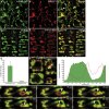
Golgi-Independent Transport of the V-ATPase and the V-PPase AVP1/VHP1 to the Tonoplast. (A) to (G) CLSM of cortex cells in the wild type (A) and gnl1;GNL1-BFAS ([B] to [G]). (A) In cells expressing VHA-a3-GFP and VMA12-RFP, VHA-a3-GFP cannot be detected in the ER. (B) In untreated cells, VHA-a1-GFP labels the TGN/EE, whereas VHA-a3-mRFP is found at the tonoplast. (C) After 3 h in the presence of ConcA, VHA-a1-GFP and VHA-a3-mRFP do not colocalize. (D) After 3 h in the presence of BFA, VHA-a1-GFP is retained in the ER while VHA-a3-mRFP is not. (E) to (G) FRAP experiment showing that in the presence of BFA, VHA-a3-mRFP is newly synthesized but does not accumulate in the ER. Bars = 10 µm. (E) Time course of fluorescence recovery for VHA-a1-GFP (green lines) and VHA-a3-mRFP (red lines) in the presence of BFA (solid lines) or BFA and CHX (dashed lines). (F) and (G) CLSM images showing the region of interest (dashed box) before, immediately after, and 200’ after photobleaching in the presence of BFA (F) or BFA + CHX (G).
Figure 2.
Figure 2.
Quantitative Analysis of AVP1/VHP1 Distribution in the Endomembrane System. (A) to (C) Immunogold EM detects VHA-a3-GFP ([A], arrowheads) and AVP1/VHP1 ([B] and [C]) at the tonoplast. (D) AVP1/VHP1 is detected at the tonoplast but not in the limiting membrane of MVBs (arrowheads). (E) and (F) Upon 12 h treatment with ConcA, AVP1/VHP1 is not detected in Golgi stacks or TGN-derived elements despite an unchanged amount of signal at the tonoplast. G, Golgi; V, vacuole. Bars = 400 nm in (A) to (F). (G) Quantification of labeling density (gold particles per micrometer of membrane) at vacuoles (VAC), provacuoles (PRV), MVB, TGN, Golgi, ER, and plastids/mitochondria (P/M). Error bars represent the sd for n = 30.
Figure 3.
Figure 3.
Sterol Enrichment at the Tonoplast Occurs Independently of Transport via the Golgi. (A) Filipin-sterol fluorescence colocalizes with VHA-a3-GFP (arrow) in young cells lacking a large central vacuole. (B) Filipin-sterol signal colocalizes with VHAa3-GFP at the tonoplast in elongated cells. (C) Ultrastructural analysis reveals 20- to 30-nm membrane deformations (arrowheads) typical of filipin-sterol complexes at the tonoplast of young and mature vacuoles (left) but absent from vacuoles of samples treated with DMSO solvent (right). V, vacuole. (D) In Col-0 seedlings treated with BFA for 2 h, filipin-sterol fluorescence colabels with the cytokinesis-related syntaxin KNOLLE (KN) in BFA bodies, but also stains additional membranes that are neither part of the cell plate nor of BFA bodies (arrows). (E) In gnl1 treated with BFA (top panels), while KN is retained in the ER, filipin-sterol fluorescence mostly highlights membranes distinct from the ER (arrows). In untreated gnl1 (bottom panels), colabeling between KN and sterols was observed. (F) In gnl1/GNL1-BFAS treated with BFA (top panels), while KN is retained in the ER, filipin-sterol fluorescence mostly highlights membranes distinct from the ER. In untreated gnl1/GNL1-BFAS (bottom panels), colabeling between KN and sterols was observed. Bars = 10 µm in (A), (B), and (D) to (F) and 400 nm in (C).
Figure 4.
Figure 4.
Vacuole Morphology in the Root Meristem. (A) to (C) EM of high-pressure frozen root tips. (A) Vacuoles of variable size can be detected in all cells of the root meristem including the quiescent center (asterisks). (B) Meristematic cell with multiple small vacuoles, some of which appear tubular. (C) Vacuoles are also present in meristematic cells during cytokinesis. (D) Root meristem stained with FM4-64 (red) and BCECF (green); vacuoles can be detected in all cell types with the exception of stele and central columella cells. (E) to (H) Projection of Z-stacks ([E] and [G]) and surface renderings ([F] and [H]) show that each cell contains a single complex tubular network that is equally distributed among the two daughter cells during cytokinesis ([G] and [H]). Bars = 4 µm in (A), 1 µm in (B) and (C), 20 µm in (D) and (E), and 5 µm in (G).
Figure 5.
Figure 5.
Early Stages of Vacuole Formation. (A) to (C) Developing vacuoles in stele cells visualized by combining VHA-a3-GFP (A) with luminal staining by SNARF-1 (B). Arrowheads point to structures positive for VHA-a3-GFP but lacking luminal staining (C). (D) to (F) Developing vacuoles in cortex cells visualized by combining VHA-a3-mRFP (E) with luminal staining by BCECF (D). Arrowheads point to loop structures positive for VHA-a3-mRFP but lacking luminal staining (F). (G) Quantification of VHA-a3-mRFP loops with low luminal BCECF staining in cells of the meristematic and elongation zones. Error bars indicate the sd for n = 10 roots. (H) Fluorescence intensity profile of a loop and a vacuole (white line) indicating that VHA-a3-mRFP fluorescence in loops is twofold higher than in the neighboring tonoplast. (I) Time-lapse imaging showing the conversion of a loop labeled by VHA-a3-mRFP to a tubular structure filled with BCECF (see Supplemental Movie 3 online). Bars = 10 µm in (A) to (F) and (H) and 5 µm in (I).
Figure 6.
Figure 6.
Provacuoles Are Double-Membrane Structures Characterized by the Presence of V-ATPase and V-PPase. (A) EM of a circular structure consisting of two closely apposed membranes that seemingly engulfs a portion of cytosol. (B) to (E) Immunogold cytochemistry of ultrathin sections after high-pressure freezing and freeze substitution. (B) Consecutive ultrathin sections show AVP1/VHP1 labeling associated with 30-nm-thick double bilayered provacuole (see inset magnification, right). CP, cell plate. (C) A semicircular provacuole showing AVP1/VHP1 labeling (inset) with an expanding lumen toward the edges. (D) AVP1/VHP1 uniformly labels a developing vacuole displaying consecutive constrictions (inset) and enlargements of its lumen. (E) Anti-GFP immunogold labeling of VHA-a3-GFP revealing its presence on provacuoles. (F) Constrictions that separate two adjacent lumina represent an additional feature of provacuoles. Bars = 400 nm in (A) to (F) and 60 nm in magnification inset of (B) (right).
Figure 7.
Figure 7.
Provacuoles Do Not Originate from Post-Golgi Trafficking and Are Distinct from Autophagosomes. (A) to (C) Quantification of loops in the wild type and pat2-2 showing that pat2-2 has more loops with a higher fluorescence intensity in the meristematic zone (B) as well as in the elongation zone (C). Error bars for the number of loops in (A) represent the sd for n = 10 roots and for the fluorescence intensity of n = 65 loops. (D) to (I) Immunogold cytochemistry of AVP1/VHP1 on ultrathin sections after high-pressure freezing and freeze substitution. (D) Ultrastructural analysis of the pat2-1 mutant. Provacuoles are present but display an aberrant multilayered morphology. (E) Ultrastructural analysis of a VPS45-silenced line (siVPS45). Meristematic root cells contain multilayered provacuoles. (F) Treatment with ConcA induces formation of multilayered provacuoles after 1 h of treatment. (G) Upon 6 h of BFA treatment of the gnl1 mutant, provacuoles were still present. (H) In the amsh3 mutant, morphologically aberrant provacuoles were present, sometimes with multilayered profiles. (I) In the BFA-treated gnl1 mutants, double membrane structures without immunogold labeling are observed next to AVP1-labeled provacuoles. (J) In the atg2-1 mutant defective in autophagosome formation, provacuoles of normal morphology are present. (K) and (L) Confocal microscopy of seedlings expressing GFP-ATG8b stained with SNARF-1. Autophagosomes labeled with GFP-ATG8b are distinct from the acidified vacuolar lumen (K) and do not accumulate on provacuolar loops (L). Rectangles indicate magnified areas in (E), (F), (J), and (I). Bars = 10 µm in (B), (C), and (K), 1 µm in (L), 400 nm in (D) to (H) and (J), and 200 nm in (I).
Figure 8.
Figure 8.
Do Provacuoles Originate from the ER? (A) and (B) CLSM codetection of VHA-a3-mRFP and its assembly factor VMA21-GFP in root cortex cells reveals lack of colocalization in the ER and small vacuoles devoid of VMA21-GFP at the periphery of the nuclear envelope. (C) Provacuole connected to the cortical ER as observed by immunogold EM with anti-AVP1/VHP1 after ConcA treatment. (D) Schematic model of vacuolar biogenesis. V-ATPase assembly takes place in the ER (I). V-ATPase and V-PPase are sorted into a subdomain of the smooth ER that differs in lipid composition (II). The resulting double-membrane sheet curves into a cup-shaped structure with minimal luminal volume (III). Increase in luminal volume leads to formation of ball-shaped or tubular structures (IV). Fusion with preexisting vacuoles can occur at any stage (V). Bars = 10 µm in (A) and (B) and 400 nm in (C).

References

    1. Amelunxen F., Heinze U. (1984). Zur entwicklung der vacuole in testa-zellen des leinsamens. Eur. J. Cell Biol. 35: 343–354
    1. Bassham D.C., Sanderfoot A.A., Kovaleva V., Zheng H., Raikhel N.V. (2000). AtVPS45 complex formation at the trans-Golgi network. Mol. Biol. Cell 11: 2251–2265 - PMC - PubMed
    1. Berjak P. (1972). Lysosomal compartmentation: Ultrastructural aspects of the origin, development, and function of vacuoles in root cells of Lepidium sativum. Ann. Bot. (Lond.) 36: 73–81
    1. Boutté Y., Frescatada-Rosa M., Men S., Chow C.-M., Ebine K., Gustavsson A., Johansson L., Ueda T., Moore I., Jürgens G., Grebe M. (2010). Endocytosis restricts Arabidopsis KNOLLE syntaxin to the cell division plane during late cytokinesis. EMBO J. 29: 546–558 - PMC - PubMed
    1. Boutté Y., Grebe M. (2009). Cellular processes relying on sterol function in plants. Curr. Opin. Plant Biol. 12: 705–713 - PubMed

Publication types

MeSH terms