. 2013 Sep;25(9):3389-404.
doi: 10.1105/tpc.113.114736. Epub 2013 Sep 6.

Arabidopsis kinetochore null2 is an upstream component for centromeric histone H3 variant cenH3 deposition at centromeres

Affiliations

Arabidopsis kinetochore null2 is an upstream component for centromeric histone H3 variant cenH3 deposition at centromeres

Inna Lermontova et al. Plant Cell. 2013 Sep.

Abstract

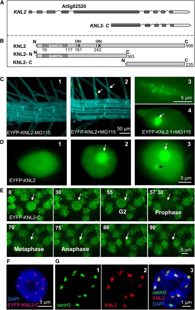
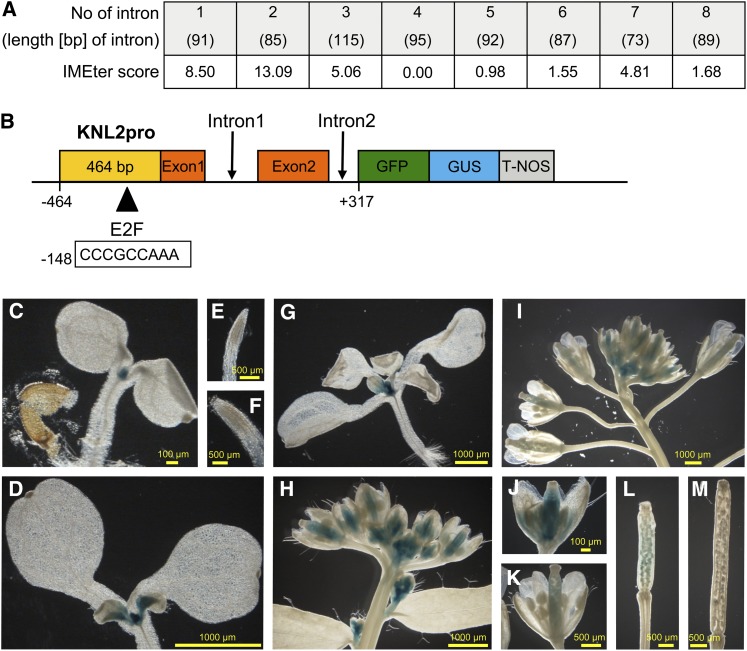
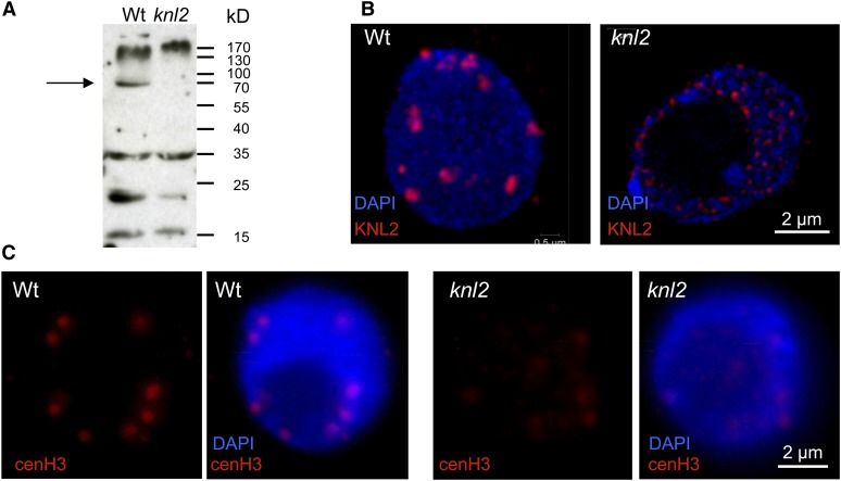
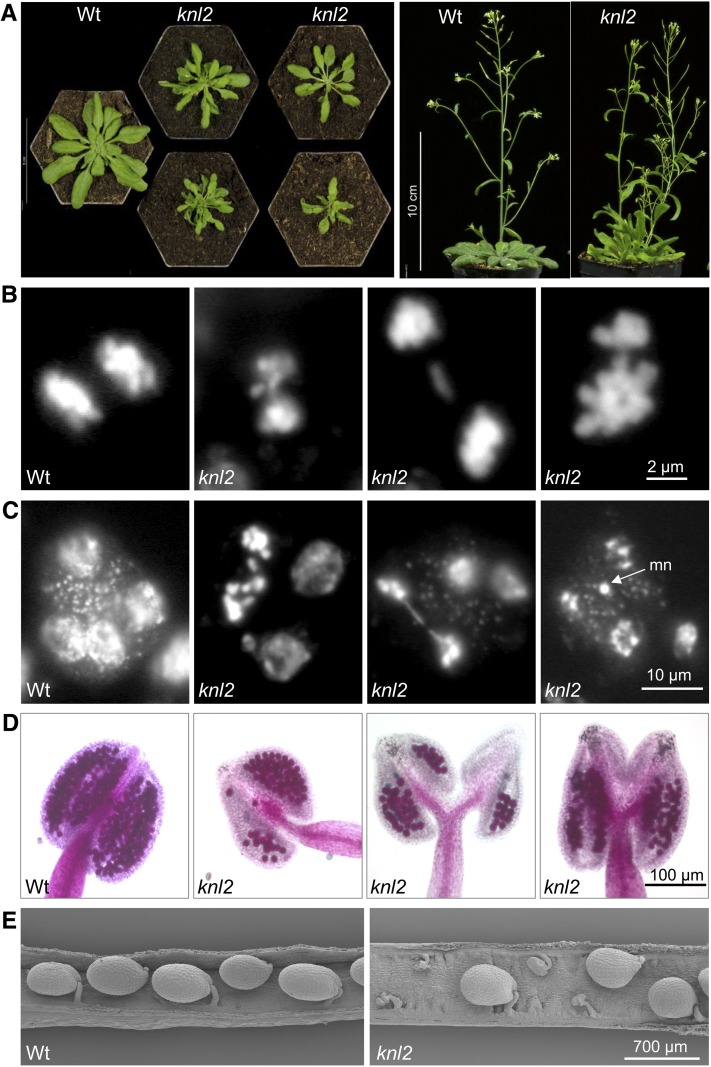
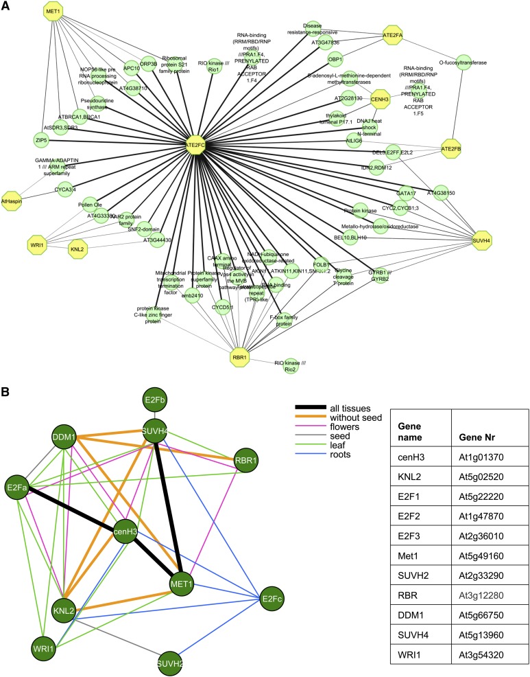
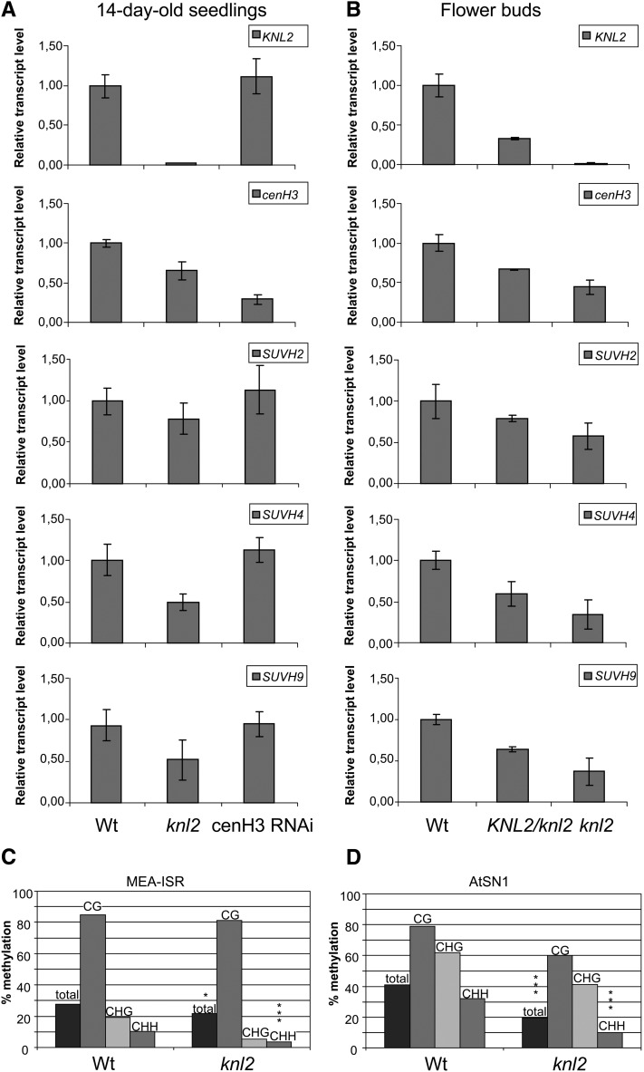
The centromeric histone H3 variant cenH3 is an essential centromeric protein required for assembly, maintenance, and proper function of kinetochores during mitosis and meiosis. We identified a kinetochore null2 (KNL2) homolog in Arabidopsis thaliana and uncovered features of its role in cenH3 loading at centromeres. We show that Arabidopsis KNL2 colocalizes with cenH3 and is associated with centromeres during all stages of the mitotic cell cycle, except from metaphase to mid-anaphase. KNL2 is regulated by the proteasome degradation pathway. The KNL2 promoter is mainly active in meristematic tissues, similar to the cenH3 promoter. A knockout mutant for KNL2 shows a reduced level of cenH3 expression and reduced amount of cenH3 protein at chromocenters of meristematic nuclei, anaphase bridges during mitosis, micronuclei in pollen tetrads, and 30% seed abortion. Moreover, knl2 mutant plants display reduced expression of suppressor of variegation 3-9 homologs2, 4, and 9 and reduced DNA methylation, suggesting an impact of KNL2 on the epigenetic environment for centromere maintenance.


Figures

Figure 1.
Figure 1.
KNL2 Gene and Protein Structures and Subcellular Localization of KNL2. (A) Structure of KNL2 gene and the part of the gene encoding the C-terminal part of KNL2. Exons are shown as dark-gray boxes. (B) Schematic of KNL2 protein structure (top fragment). The conserved protein domain SANTA (amino acids 19 to aa117) and putative ubiquitination sites at positions 161 and 242 are indicated. KNL2-N (middle fragment) and KNL2-C (bottom fragment) were separately fused with EYFP for localization studies. KNL2-C was used for expression of recombinant protein. (C) Roots of EYFP-KNL2 transgenic plants without (1) and with (2) treatment with the proteasome inhibitor MG115. Expression of EYFP-KNL2 was detected only after treatment with MG115. KNL2 is detectable in nucleoplasm and at chromocenters (3) or in nucleoplasm and nucleolus (white arrow) and weak at chromocenters (4). (D) Transient expression of the EYFP-KNL2 fusion construct in N. benthamiana. Three compartments are labeled: nucleoplasm (1), nucleoplasm and nucleoli (2), and nucleoplasm, nucleoli, and nuclear bodies (3). (E) Live imaging of root tip cells of Arabidopsis transformed with the EYFP-KNL-C fusion construct. A cell undergoing mitosis is indicated by an arrow. (F) Nucleus of an EYFP-KNL2-C transformed Arabidopsis plant immunostained with anti-GFP antibodies showed colocalization of EYFP-cenH3 immunosignals with bright DAPI-stained chromocenters. (G) Double immunostaining of a meristematic nucleus of an EYFP-cenH3 transformant with anti-GFP for the EYFP-cenH3 (1) and anti-KNL2 antibodies (2). Colocalization of immunosignals for both proteins (3). In (F) and (G), (3) DNA is counterstained with DAPI.
Figure 2.
Figure 2.
KNL2pro Activity in Arabidopsis during Development. (A) IMEter scores, indicating predicted intron mediated enhancement of transcription, for all introns of KNL2 as determined by in silico analysis using the IMEter online program (Rose et al., 2008). (B) Scheme of the KNL2pro:GFP-GUS reporter gene construct. (C) to (M) Histochemical localization of GUS activity in Arabidopsis plants transgenic for AtKNL2pro:GFP-GUS: 4-d-old (C) and 7-d-old (D) seedling as well as their primary root tips ([E] and [F]). Fourteen-day-old seedling (G), inflorescences ([H] and [I]), mature flowers ([J] and [K]), and siliques ([L] and [M]) at different developmental stages.
Figure 3.
Figure 3.
KNL2 and cenH3 Protein Levels in Atknl2 Mutant Compared with the Wild Type. (A) Immunoblot analysis with anti-KNL2 antibodies on protein extracts isolated from seedlings of Arabidopsis wild-type (Wt) and knl2 T-DNA insertion mutants. Arrow shows the position of KNL2 protein (∼66 kD) in the wild type and its absence in the knl2 mutant. (B) Immunostaining of meristematic nuclei of Arabidopsis wild type and the knl2 mutant with anti-KNL2 antibodies. (C) Immunostaining of meristematic nuclei of Arabidopsis wild type and knl2 mutant with anti-cenH3 antibodies. Immunostaining experiment was repeated three times, and each time 10 root tips of the wild type and knl2 mutant were analyzed. In (B) and (C), DNA is counterstained with DAPI.
Figure 4.
Figure 4.
Phenotype of the knl2 Mutant. (A) Comparison of the phenotype of knl2 mutant with that of the wild-type (Wt). Plants were grown for 4 weeks (left panel) or 6 weeks (right panel) on soil. (B) Mitotic anaphases of the wild type and of a knl2 mutant plant with bridges and lagging chromosomes. Among 400 anaphases of the wild type and 1500 anaphases of the knl2 mutant, 0.5 and 10% showed bridges, respectively. (C) Pollen tetrads of the wild type and of a knl2 mutant with bridges and micronuclei (mn), respectively. (D) Anthers of the wild type and of a knl2 mutant after Alexander staining to indicate viable pollen. (E) Scanning electron microscopy images of siliques of the wild type and a knl2 mutant.
Figure 5.
Figure 5.
Gene Regulatory Network for cenH3 Deposition in Arabidopsis and Coexpression of KNL2 and cenH3 with Genes of the RBR-E2F Transcription Regulation Pathway and with DNA and H3 Methyltransferases. (A) Key regulator genes are indicated by yellow octagons. The network is inferred using collected gene expression data from the National Center for Biotechnology Information Gene Expression Omnibus (see Methods). The edge width represents the interaction score calculated by MRNET algorithm (Meyer et al., 2008). KNL2, kinetochore null2; cenH3, centromeric histone H3; MET1, methyltransferase 1; E2Fa, E2Fb, E2Fc, Arabidopsis thaliana E2F transcription factors; RBR1, retinoblastoma related1; SUVH4, histomemethyltransferase; Haspin, Haspin kinase; WRI1, sequence-specific DNA binding transcription factor WRINKLED1. (B) Tissue-specific coexpression patterns are derived from predefined microarray sets in the CORNET program. Different colors indicate the tissues that display a significant correlation of expression between the corresponding genes linked by a bar (Pearson coefficient at least 0.6 and P value < 0.05).
Figure 6.
Figure 6.
Expression of KNL2, cenH3, SUVH2, SUVH4, and SUVH9 in the knl2 Mutant and cenH3 RNAi Plants and DNA Methylation at MEA-ISR and At-SN1 Loci in knl2 Mutants. (A) and (B) Real-time quantitative RT-PCR analysis of KNL2, cenH3, SUVH2, SUVH4, and SUVH9 mRNA transcripts in 14-d-old seedlings of the knl2 mutant and cenH3 RNAi transformants compared with the wild type (A) and in flower buds of heterozygous KNL2/knl2 and homozygous knl2 mutants compared with the wild type (B). Amplification of ACTIN2 mRNA was used for data normalization. (C) and (D) DNA methylation was determined by bisulfite sequencing of genomic DNA of 7-d-old seedlings. Individual clones were sequenced for the different genotypes. The bars mark the levels of DNA methylation in percentage of methylated cytosines relative to total cytosines. Wt, wild-type plants (Columbia-0). (C) DNA methylation at MEA-ISR. Significant differences in total DNA methylation between the different genotypes were checked by χ2 tests. Wild type, n = 26; knl2, n = 19 (complete methylation: χ2, 5.54, *P < 0.05; asymmetric methylation: χ2, 18.24, ***P < 0.001). (D) DNA methylation at At-SN1. Wild type, n = 12; At-knl2, n = 10 (χ2, 45.48, ***P < 0.001; asymmetric methylation: χ2, 51.30, ***P < 0.001).

Similar articles

Cited by

References

    1. Aasland R., Stewart A.F., Gibson T. (1996). The SANT domain: A putative DNA-binding domain in the SWI-SNF and ADA complexes, the transcriptional co-repressor N-CoR and TFIIIB. Trends Biochem. Sci. 21: 87–88 - PubMed
    1. Alexander M.P. (1969). Differential staining of aborted and nonaborted pollen. Stain Technol. 44: 117–122 - PubMed
    1. Allshire R.C. (1997). Centromeres, checkpoints and chromatid cohesion. Curr. Opin. Genet. Dev. 7: 264–273 - PubMed
    1. Barnhart M.C., Kuich P.H., Stellfox M.E., Ward J.A., Bassett E.A., Black B.E., Foltz D.R. (2011). HJURP is a CENP-A chromatin assembly factor sufficient to form a functional de novo kinetochore. J. Cell Biol. 194: 229–243 - PMC - PubMed
    1. Berckmans B., De Veylder L. (2009). Transcriptional control of the cell cycle. Curr. Opin. Plant Biol. 12: 599–605 - PubMed

Publication types

MeSH terms

Associated data