Navigated DENSE strain imaging for post-radiofrequency ablation lesion assessment in the swine left atria
- PMID: 24014803
- PMCID: PMC3924042
- DOI: 10.1093/europace/eut229
Navigated DENSE strain imaging for post-radiofrequency ablation lesion assessment in the swine left atria
Abstract
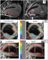
Aims: Prior work has demonstrated that magnetic resonance imaging (MRI) strain can separate necrotic/stunned myocardium from healthy myocardium in the left ventricle (LV). We surmised that high-resolution MRI strain, using navigator-echo-triggered DENSE, could differentiate radiofrequency ablated tissue around the pulmonary vein (PV) from tissue that had not been damaged by radiofrequency energy, similarly to navigated 3D myocardial delayed enhancement (3D-MDE).
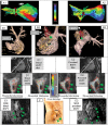
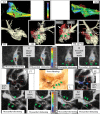
Methods and results: A respiratory-navigated 2D-DENSE sequence was developed, providing strain encoding in two spatial directions with 1.2 × 1.0 × 4 mm(3) resolution. It was tested in the LV of infarcted sheep. In four swine, incomplete circumferential lesions were created around the right superior pulmonary vein (RSPV) using ablation catheters, recorded with electro-anatomic mapping, and imaged 1 h later using atrial-diastolic DENSE and 3D-MDE at the left atrium/RSPV junction. DENSE detected ablation gaps (regions with >12% strain) in similar positions to 3D-MDE (2D cross-correlation 0.89 ± 0.05). Low-strain (<8%) areas were, on average, 33% larger than equivalent MDE regions, so they include both injured and necrotic regions. Optimal DENSE orientation was perpendicular to the PV trunk, with high shear strain in adjacent viable tissue appearing as a sensitive marker of ablation lesions.
Conclusions: Magnetic resonance imaging strain may be a non-contrast alternative to 3D-MDE in intra-procedural monitoring of atrial ablation lesions.
Keywords: Atrial fibrillation; Magnetic resonance imaging ablation; Porcine model.
Figures


Similar articles
-
Acute enhancement of necrotic radio-frequency ablation lesions in left atrium and pulmonary vein ostia in swine model with non-contrast-enhanced T1 -weighted MRI.Magn Reson Med. 2020 Apr;83(4):1368-1379. doi: 10.1002/mrm.28001. Epub 2019 Sep 30. Magn Reson Med. 2020. PMID: 31565818 Free PMC article.
-
Electroanatomic mapping and radiofrequency ablation of porcine left atria and atrioventricular nodes using magnetic resonance catheter tracking.Circ Arrhythm Electrophysiol. 2009 Dec;2(6):695-704. doi: 10.1161/CIRCEP.109.882472. Circ Arrhythm Electrophysiol. 2009. PMID: 19841033
-
Detection of pulmonary vein and left atrial scar after catheter ablation with three-dimensional navigator-gated delayed enhancement MR imaging: initial experience.Radiology. 2007 Jun;243(3):690-5. doi: 10.1148/radiol.2433060417. Radiology. 2007. PMID: 17517928 Clinical Trial.
-
Real-time magnetic resonance imaging-guided radiofrequency atrial ablation and visualization of lesion formation at 3 Tesla.Heart Rhythm. 2011 Feb;8(2):295-303. doi: 10.1016/j.hrthm.2010.10.032. Epub 2010 Oct 27. Heart Rhythm. 2011. PMID: 21034854 Free PMC article.
-
In vitro photoacoustic visualization of myocardial ablation lesions.Heart Rhythm. 2014 Jan;11(1):150-7. doi: 10.1016/j.hrthm.2013.09.071. Epub 2013 Sep 27. Heart Rhythm. 2014. PMID: 24080065 Free PMC article.
Cited by
-
Quantitative tissue-tracking cardiac magnetic resonance (CMR) of left atrial deformation and the risk of stroke in patients with atrial fibrillation.J Am Heart Assoc. 2015 Apr 27;4(4):e001844. doi: 10.1161/JAHA.115.001844. J Am Heart Assoc. 2015. PMID: 25917441 Free PMC article.
-
Cardiovascular magnetic resonance imaging of scar development following pulmonary vein isolation: a prospective study.PLoS One. 2014 Sep 24;9(9):e104844. doi: 10.1371/journal.pone.0104844. eCollection 2014. PLoS One. 2014. PMID: 25251403 Free PMC article.
-
Impact of ablation on regional strain from 4D computed tomography in the left atrium.J Interv Card Electrophysiol. 2025 Jun 20. doi: 10.1007/s10840-025-02087-8. Online ahead of print. J Interv Card Electrophysiol. 2025. PMID: 40540199
-
Measuring Cardiac Dyssynchrony with DENSE (Displacement Encoding with Stimulated Echoes)-A Systematic Review.Rev Cardiovasc Med. 2023 Sep 18;24(9):261. doi: 10.31083/j.rcm2409261. eCollection 2023 Sep. Rev Cardiovasc Med. 2023. PMID: 39076380 Free PMC article.
-
MRI use for atrial tissue characterization in arrhythmias and for EP procedure guidance.Int J Cardiovasc Imaging. 2018 Jan;34(1):81-95. doi: 10.1007/s10554-017-1179-y. Epub 2017 Jun 7. Int J Cardiovasc Imaging. 2018. PMID: 28593399 Free PMC article. Review.
References
-
- Fuster V, Rydén LE, Cannom DS, Crijns HJ, Curtis AB, Ellenbogen KA, et al. ACC/AHA/ESC 2006 Guidelines for the Management of Patients with Atrial Fibrillation: a report of the American College of Cardiology/American Heart Association Task Force on Practice Guidelines and the European Society of Cardiology Committee for Practice Guidelines. Circulation. 2006;114:e257–354. - PubMed
-
- Calkins H, Brugada J, Packer DL, Cappato R, Chen SA, Crijns HJ, et al. HRS/EHRA/ECAS expert Consensus Statement on catheter and surgical ablation of atrial fibrillation: recommendations for personnel, policy, procedures and follow-up. A report of the Heart Rhythm Society (HRS) Task Force on catheter and surgical ablation of atrial fibrillation. Heart Rhythm. 2007;4:816–61. - PubMed
-
- Cheema A, Dong J, Dalal D, Marine JE, Henrikson CA, Spragg D, et al. Incidence and time course of early recovery of pulmonary vein conduction after catheter ablation of atrial fibrillation. J Cardiovasc Electrophysiol. 2007;18:387–91. - PubMed
-
- Cheema A, Dong J, Dalal D, Vasamreddy CR, Marine JE, Henrikson CA, et al. Long-term safety and efficacy of circumferential ablation with pulmonary vein isolation. J Cardiovasc Electrophysiol. 2006;17:1080–5. - PubMed
-
- Badger TJ, Adjei-Poku YA, Burgon NS, Kalvaitis S, Shaaban A, Sommers DN, et al. Initial experience of assessing esophageal tissue injury and recovery using delayed-enhancement MRI after atrial fibrillation ablation. Circ Arrhythm Electrophysiol. 2009;2:620–5. - PubMed
Publication types
MeSH terms
Grants and funding
LinkOut - more resources
Full Text Sources
Other Literature Sources
Medical

