. 2013 Oct 25;113(10):1117-27.
doi: 10.1161/CIRCRESAHA.113.301306. Epub 2013 Sep 6.

MicroRNA-663 regulates human vascular smooth muscle cell phenotypic switch and vascular neointimal formation

Affiliations

MicroRNA-663 regulates human vascular smooth muscle cell phenotypic switch and vascular neointimal formation

Pan Li et al. Circ Res. .

Abstract

Rationale: Abnormal phenotypic switch of vascular smooth muscle cell (VSMC) is a hallmark of vascular disorders such as atherosclerosis and restenosis after angioplasty. MicroRNAs (miRNAs) have emerged as important regulators for VSMC function, and we recently identified miR-663 as critical for controlling human aortic smooth muscle cell proliferation.

Objective: To investigate whether miR-663 plays a role in human VSMC phenotypic switch and the development of neointima formation.

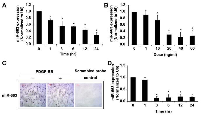
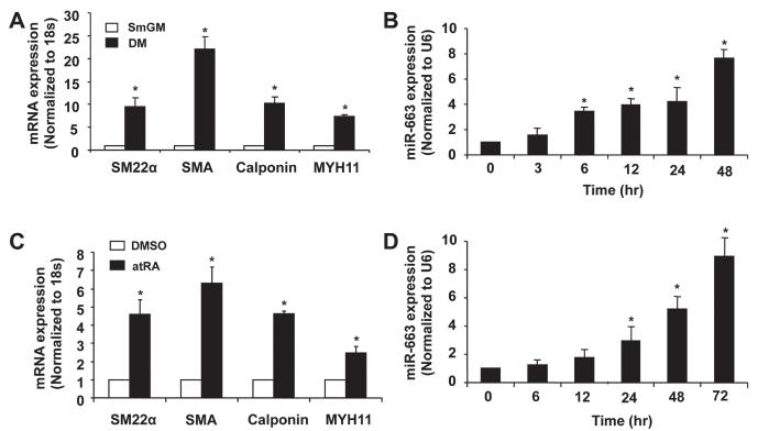
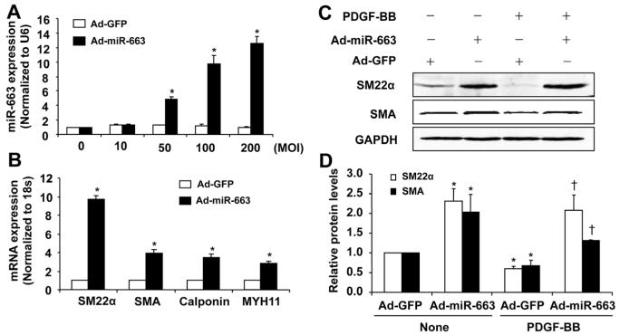
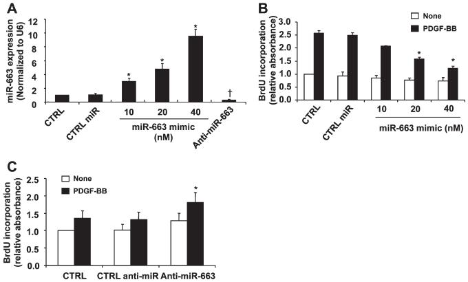
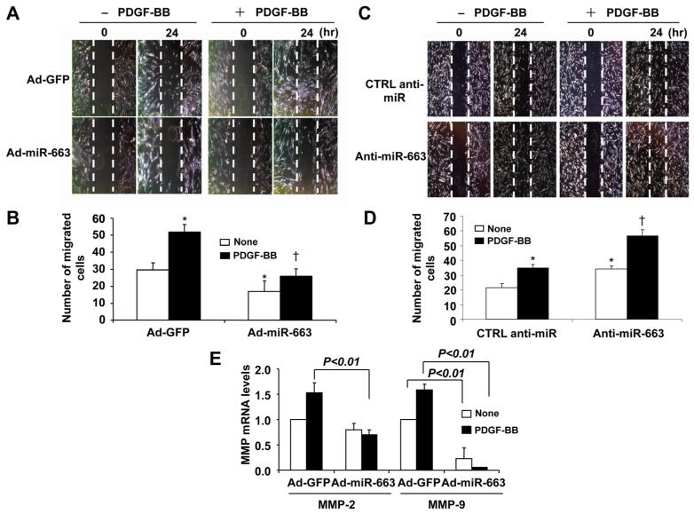
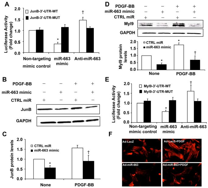
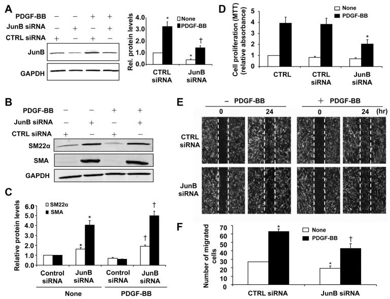
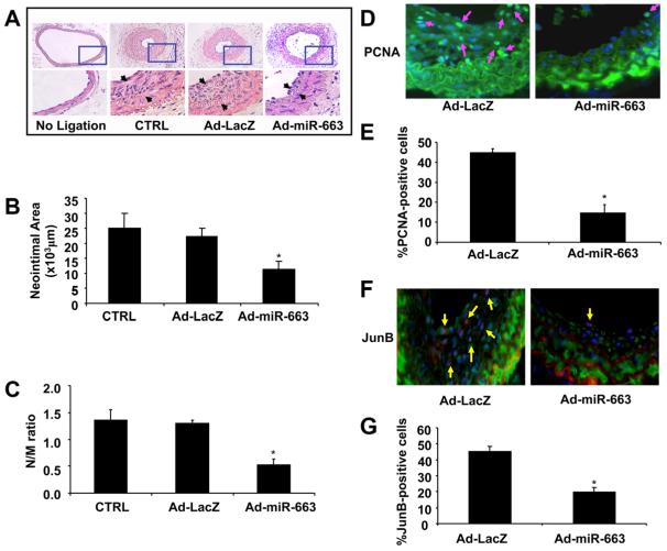
Methods and results: By using quantitative reverse-transcription polymerase chain reaction, we found that miR-663 was significantly downregulated in human aortic VSMCs on platelet-derived growth factor treatment, whereas expression was markedly increased during VSMC differentiation. Furthermore, we demonstrated that overexpression of miR-663 increased expression of VSMC differentiation marker genes, such as smooth muscle 22α, smooth muscle α-actin, calponin, and smooth muscle myosin heavy chain, and potently inhibited platelet-derived growth factor-induced VSMC proliferation and migration. We identified the transcription factor JunB and myosin light chain 9 as downstream targets of miR-663 in human VSMCs, because overexpression of miR-663 markedly inhibited expression of JunB and its downstream molecules, such as myosin light chain 9 and matrix metalloproteinase 9. Finally, we showed that adeno-miR-663 markedly suppressed the neointimal lesion formation by ≈50% in mice after vascular injury induced by carotid artery ligation, specifically via decreased JunB expression.

Conclusions: These results indicate that miR-663 is a novel modulator of human VSMC phenotypic switch by targeting JunB/myosin light chain 9 expression. These findings suggest that targeting miR-663 or its specific downstream targets in human VSMCs may represent an attractive approach for the treatment of proliferative vascular diseases.

Keywords: miR-663; migration; proliferation; vascular remodeling; vascular smooth muscle cells.


Figures

Figure 1
Figure 1. MicroRNA (miR)-663 is significantly downregulated in proliferating vascular smooth muscle cells (VSMCs)
A, Platelet-derived growth factor-BB (PDGF-BB; 20 ng/mL) caused a time-dependent decrease in miR-663 expression in human aortic VSMCs, as determined by quantitative reverse-transcription polymerase chain reaction (qRT-PCR) (n=4). *P<0.05 compared with 0 h group. B, PDGF-BB caused a dose-dependent decrease in miR-663 expression in human aortic VSMCs at 24 h after PDGF treatment, as determined by qRT-PCR (n=4). *P<0.05 compared with that in VSMCs without PDGF-BB. C, In situ hybridization (violet) shows miR-663 was significantly decreased in PDGF-BB–stimulated (20 ng/mL) VSMCs. D, Serum (5%) caused a time-dependent decrease in miR-663 expression in human aortic VSMCs, as demonstrated by qRT-PCR (n=4). *P<0.05 compared with that without 5% fetal bovine serum treatment.
Figure 2
Figure 2. MicroRNA (miR)-663 is upregulated in differentiated human aortic smooth muscle cells (SMCs)
A, Vascular SMCs (VSMCs) cultured in SMC differentiation medium (DM) for 48 h caused a significantly increased expression of VSMC differentiation marker genes, such as smooth muscle α-actin, smooth muscle 22α, calponin, and smooth muscle myosin heavy chain, as determined by quantitative reverse-transcription polymerase chain reaction (qRT-PCR) (n=5).*P<0.05 compared with SMC growth medium (smGM). B, Human aortic SMCs were cultured in SMC differentiation medium to induce SMC differentiation and the expression of miR-663 was measured by qRT-PCR at different time points (n=5). *P<0.05 compared with 0 h group. C, Human aortic SMCs were treated with all transretinoic acid (atRA; 1 mmol/L) or dimethyl sulfoxide (DMSO) and the expression of SMC differentiation marker genes was determined by qRT-PCR after 72 h of atRA treatment (n=5). *P<0.05 compared with control D, miR-663 was significantly upregulated in atRA-treated VSMCs, as determined by qRT-PCR miRNA assay (n=5). *P<0.05 compared with 0 h.
Figure 3
Figure 3. MicroRNA (miR)-663 regulates the expression of vascular smooth muscle cell (VSMC) contractile genes
A, Adenovirus-mediated overexpression of miR-663 (Ad-miR-663) with a multiplicity of infection (MOI) of 100 increases miR-663 levels in a dose-dependent manner (n=4). *P<0.05 vs control viruses expressing green fluorescent protein (Ad-GFP). B, Seventy-two hours after transduction of cell with Ad-miR-663 (MOI=100), the expression of VSMC differentiation marker genes in human aortic SMCs was determined by quantitative reverse-transcription polymerase chain reaction (qRT-PCR) (n=5). *P<0.05 compared with Ad-GFP group. C, Ad-miR-663 (MOI=100) increases the expression of SMC differentiation marker genes in human aortic SMCs in the presence and absence of platelet-derived growth factor (PDGF; 20 ng/mL) treatment, as determined by Western blot analysis. D, Densitometric analysis of SMC differentiation marker genes expression as determined by Western blot (n=5). *P<0.05 vs Ad-GFP without PDGF treatment; †P<0.05 vs Ad-GFP with PDGF (20 ng/mL) treatment.
Figure 4
Figure 4. Role of microRNAs (miR)-663 in vascular smooth muscle cell (VSMC) proliferation
A, Human aortic VSMCs were transfected with control miR mimics, miR-663 mimics, or miR-663 inhibitors, respectively, and then subjected to quantitative reverse-transcription polymerase chain reaction (qRT-PCR) analysis of miR-663 level (n=4). *P<0.05 vs CTRL miR; †P<0.05 vs control (CTRL) miR. B, miR-663 mimic inhibits platelet-derived growth factor-BB (PDGF-BB)-induced VSMC proliferation in a dose-dependent manner as measured by BrdU incorporation (n=4). *P<0.05 vs CTRL miR with PDGF treatment. C, Forty-eight hours after transfection with either anti-miR-663 (50 nmol/L) or CTRL anti-miR (50 nmol/L), SMCs were stimulated with or without PDGF-BB (2 ng/mL) for 24 h. Anti-miR-663 (50 nmol/L) significantly increased human aortic VSMCs proliferation stimulated by PDGF-BB (2 ng/mL) as determined by BrdU incorporation (n=5). *P<0.05 compared with CTRL miR treated with PDGF-BB (2 ng/mL) for 24 h.
Figure 5
Figure 5. Role of microRNA (miR)-663 in vascular smooth muscle cell (VSMC) migration
A, Forty-eight hours after transduction of adenovirus-mediated overexpression of miR-663 (Ad-miR-663; MOI=100), VSMCs were starved and cell migration was measured after platelet-derived growth factor (PDGF; 20 ng/mL) stimulation for 24 h by scratch-wound assay. Migrated cells were quantitated and the results are shown in (B). The data are means±SD of the number of migrated cells from 3 independent experiments. *P<0.05 vs control viruses expressing green fluorescent protein (Ad-GFP) without PDGF treatment; †P<0.05 vs Ad-GFP with PDGF treatment. C, Forty-eight hours after transfected with anti-miR-663 (50 nmol/L) or anti-miR control (CTRL) (50 nmol/L), smooth muscle cells (SMCs) were subjected to injury by scraping and then were stimulated with or without PDGF-BB (20 ng/mL) for 24 h. Migrated cells were quantitated using Image J software program and the results are shown in (D). The data are means±SD of the number of migrated cells from 3 independent experiments. *P<0.05 vs CTRL anti-miR without PDGF treatment; †P<0.05 vs CTRL anti-miR with PDGF treatment. E, Ad-miR-663 decreases matrix metalloproteinase 2 (MMP-2) and MMP-9 mRNA levels in human aortic SMCs as measured by quantitative reverse-transcription polymerase chain reaction (qRT-PCR) (n=5).
Figure 6
Figure 6. Identification of JunB and myosin light chain 9 (Myl9) as target genes of microRNA (miR)-663 in vascular smooth muscle cells (VSMCs)
A, miR-663 mimic (50 nmol/L) or nontargeting control (50 nmol/L) was cotransfected with the luciferase reporter carrying WT-JunB 3′-UTR or mutated JunB 3′-UTR. Forty-eight hours after transfection, renilla luciferase activities were measured (n=5). *P<0.05 vs JunB-3′-UTR-WT transfected with mimic control; †P<0.05 vs JunB-3′-UTR-WT transfected with mimic control. B, Effect of miR-663 on JunB expression in platelet-derived growth factor-BB (PDGF-BB; 20 ng/mL)-stimulated VSMCs. C, Densitometric analysis of JunB protein levels as measured by Western blot (n=5). *P<0.05 vs control (CTRL) miR without PDGF treatment; †P<0.05 vs CTRL miR with PDGF treatment. D, Effect of miR-663 on Myl9 expression in PDGF-BB (20 ng/mL)-stimulated VSMCs. Densitometric analysis of Myl9 protein levels as measured by Western blot (n=5). *P<0.05 vs CTRL miR without PDGF treatment; †P<0.05 vs CTRL miR with PDGF treatment. E, miR-663 mimic (50 nmol/L) or nontargeting control (50 nmol/L) was cotransfected with the luciferase reporter carrying WT-Myl9 3′-UTR or mutated Myl9 3′-UTR. Forty-eight hours after transfection, renilla luciferase activities were measured (n=5). *P<0.05 vs Myl9-3′-UTR-WT transfected with mimic control; †P<0.05 vs Myl9-3′-UTR-WT transfected with mimic control. F, VSMCs were transduced with either Ad-LacZ or Ad-miR-663. Forty-eight hours after transduction, SMCs were starved for 48 h and then stimulated with 25 ng/mL PDGF-BB for 20 min. Stress fiber formation was examined by staining cells with Alexa Fluor 546 phalloidin.
Figure 7
Figure 7. JunB is critically implicated in vascular smooth muscle cell (VSMC) proliferation and migration
A, Silencing of JunB by specific small interfering RNA (JunB siRNA; 100 nmol/L) reduced JunB expression as determined by Western blot. *P<0.05 vs control (CTRL) siRNA without platelet-derived growth factor (PDGF) treatment; †P<0.05 vs CTRL siRNA with PDGF (20 ng/mL) treatment. B, Silencing JunB increases the expression of SMC marker genes smooth muscle 22α (SM22α) and smooth muscle α-actin (SMA), as determined by Western blot analysis. C, Densitometric analysis of SM22α and SMA protein levels as measured by Western blot (n=5). *P<0.05 vs CTRL siRNA without PDGF treatment; †P<0.05 vs CTRL siRNA with PDGF (20 ng/mL) treatment. D, JunB knockdown attenuated PDGF-BB (20 ng/mL)-induced proliferation in human aortic SMCs as determined by MTT assay (n=4). *P<0.05 vs control siRNA with PDGF-BB treatment. E, VSMCs were transfected with JunB siRNA (100 nmol/L). Forty-eight hours after transfection, VSMCs were starved and cell migration was measured after PDGF-BB (20 ng/mL) stimulation for 24 h by scratch-wound assay. F, Quantitation of migrated cells. The data are means±SD of the number of migrated cells from 3 independent experiments. *P<0.05 vs CTRL siRNA without PDGF treatment; †P<0.05 vs CTRL siRNA with PDGF treatment.
Figure 8
Figure 8. MicroRNA (miR)-663 attenuates neointimal formation in a mouse model of carotid artery ligation
A, Adenovirus-mediated overexpression of miR-663 (Ad-miR-663) significantly reduced neointimal formation in vivo. Representative hematoxylin and eosin (H&E)-stained carotid artery slices from mouse treated with Ad-LacZ or Ad-miR-663 at 14 days after carotid artery ligation. B and C, The effect of miR-663 on vascular neointimal lesion formation in mouse carotid arteries at 14 days after ligation injury as quantitated by neointimal area and neointima/media (N/M) ratio. *P<0.05 vs Ad-LacZ or control (CTRL) D, Representative immunofluorescent staining of PCNA in mouse carotid arteries at 14 days after ligation injury. Green is the PCNA that represents proliferating cells; blue is the cell nuclear staining by DAPI. E, Quantification of PCNA-positive cells showed that fewer cells were proliferating in the injured arteries treated with Ad-miR-663 compared with Ad-LacZ-treated vessels. *P<0.05 vs Ad-LacZ. F, Representative immunofluorescent staining of JunB in mouse carotid arteries at 14 days after ligation injury. Green is smooth muscle α-actin; purple is JunB staining; and blue is nuclear staining by DAPI. G, Quantification of JunB-positive cells showed that JunB was downregulated in Ad-miR-663-treated vessels compared with Ad-LacZ-treated vessels. *P<0.05 vs Ad-LacZ.

Comment in

References

    1. Davis-Dusenbery BN, Wu C, Hata A. Micromanaging vascular smooth muscle cell differentiation and phenotypic modulation. Arterioscler Thromb Vasc Biol. 2011;31:2370–2377. - PMC - PubMed
    1. Rensen SS, Doevendans PA, van Eys GJ. Regulation and characteristics of vascular smooth muscle cell phenotypic diversity. Neth Heart J. 2007;15:100–108. - PMC - PubMed
    1. Rangrez AY, Massy ZA, Metzinger-Le Meuth V, Metzinger L. miR-143 and miR-145: molecular keys to switch the phenotype of vascular smooth muscle cells. Circ Cardiovasc Genet. 2011;4:197–205. - PubMed
    1. Owens GK, Kumar MS, Wamhoff BR. Molecular regulation of vascular smooth muscle cell differentiation in development and disease. Physiol Rev. 2004;84:767–801. - PubMed
    1. Tallquist M, Kazlauskas A. PDGF signaling in cells and mice. Cytokine Growth Factor Rev. 2004;15:205–213. - PubMed

Publication types

MeSH terms

LinkOut - more resources