Filariasis of the buccal mucosa: A diagnostic dilemma
- PMID: 24015022
- PMCID: PMC3757895
- DOI: 10.4103/0976-237X.114883
Filariasis of the buccal mucosa: A diagnostic dilemma
Abstract
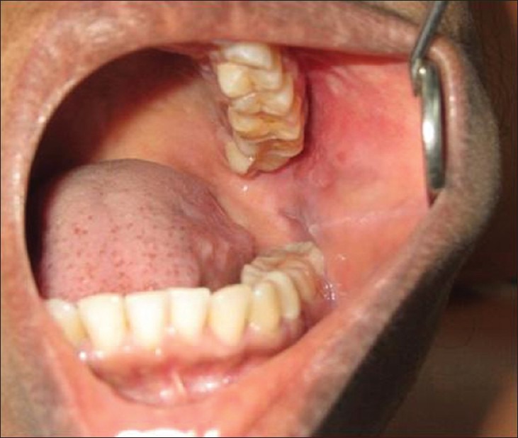
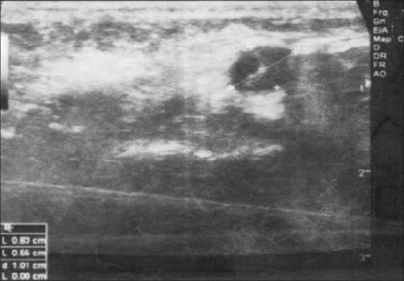
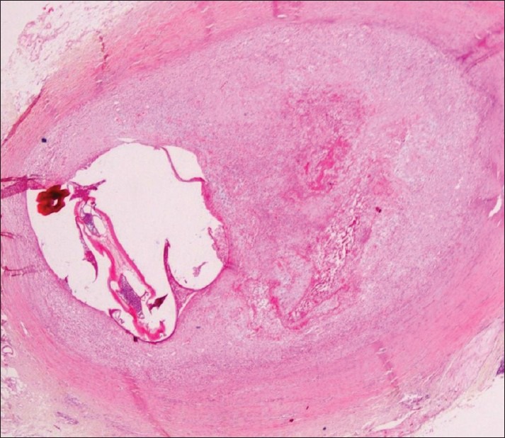
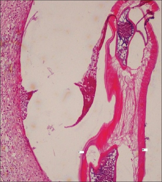
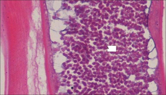
Filariasis is an endemic disease in tropical and subtropical countries. Filarial nematodes can infect humans through vectors, commonly mosquitoes. Human infection can manifest as lymphatic filariasis, subcutaneous or pulmonary nodules and with eye involvement. Intra-oral presentation is very rare and often poses a diagnostic dilemma to the dentist. We report a case of intra-oral Dirofilaria repens infection in a 54-year-old female patient, involving the buccal mucosa. History was unremarkable and on clinical examination, a diffuse swelling with no significant signs and symptoms was seen. Laboratory investigations and radiographs were non-contributory to diagnosis. Ultrasound findings revealed a hypo-echoic lesion in the muscular layer of the left cheek. Differential diagnoses considered were minor salivary gland tumor, parotid sialolith, and cysticercosis among others. The presence of a Dirofilaria worm in the excised nodule confirmed the diagnosis. Medical awareness of the risk of intra-oral nematode infection is essential. A detailed travel history, awareness of endemic status of certain diseases, proper diagnosis and management helps in better prognosis for the patient.
Keywords: Dirofilaria; filariasis; nematode infection; oral cavity.
Conflict of interest statement
Figures
References
-
- Seddon SV, Peckitt NS, Davidson RN, Sugar AW. Helminth infection of the parotid gland. J Oral Maxillofac Surg. 1992;50:183–5. - PubMed
-
- Hira PR, Al-Buloushi A, Khalid N, Iqbal J, Bain O, Eberhard ML. Zoonotic filariasis in the Arabian Peninsula: Autochthonous onchocerciasis and dirofilariasis. Am J Trop Med Hyg. 2008;79:739–41. - PubMed
-
- Sathyan P, Manikandan P, Bhaskar M, Padma S, Singh G, Appalaraju B. Subtenons infection by Dirofilaria repens. Indian J Med Microbiol. 2006;24:61–2. - PubMed
-
- Dissanaike AS, Abeyewickreme W, Wijesundera MD, Weerasooriya MV, Ismail MM. Human dirofilariasis caused by Dirofilaria (Nochtiella) repens in Sri Lanka. Parassitologia. 1997;39:375–82. - PubMed
Publication types
LinkOut - more resources
Full Text Sources
Other Literature Sources