. 2013 Aug 28;8(8):e72061.
doi: 10.1371/journal.pone.0072061. eCollection 2013.

Protein kinase C zeta regulates human pancreatic cancer cell transformed growth and invasion through a STAT3-dependent mechanism

Affiliations

Protein kinase C zeta regulates human pancreatic cancer cell transformed growth and invasion through a STAT3-dependent mechanism

Amanda M Butler et al. PLoS One. .

Abstract

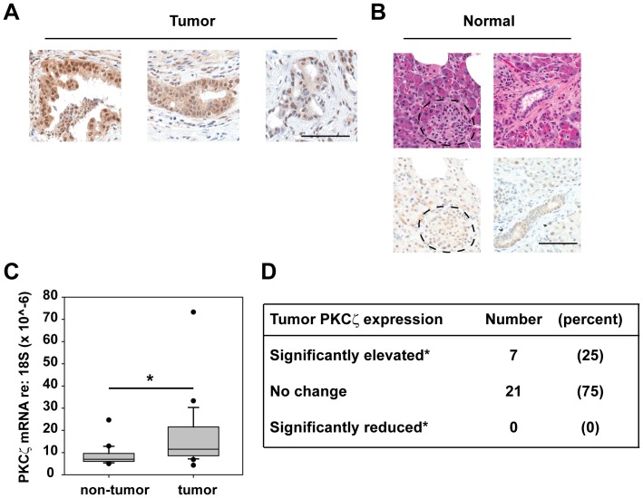
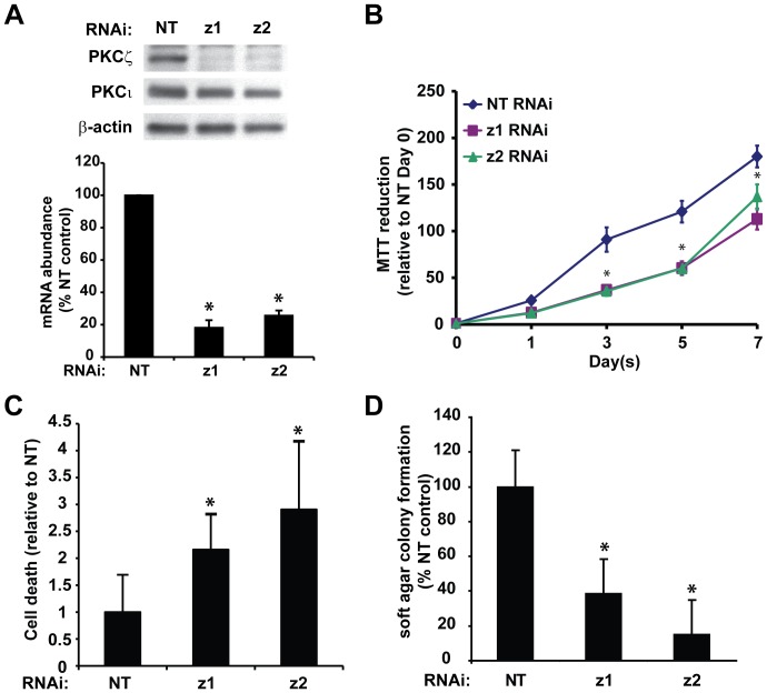
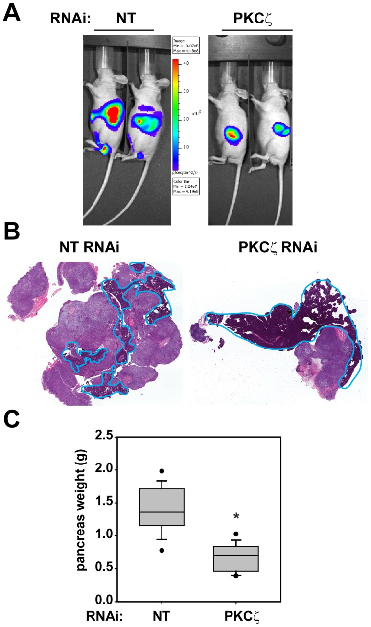
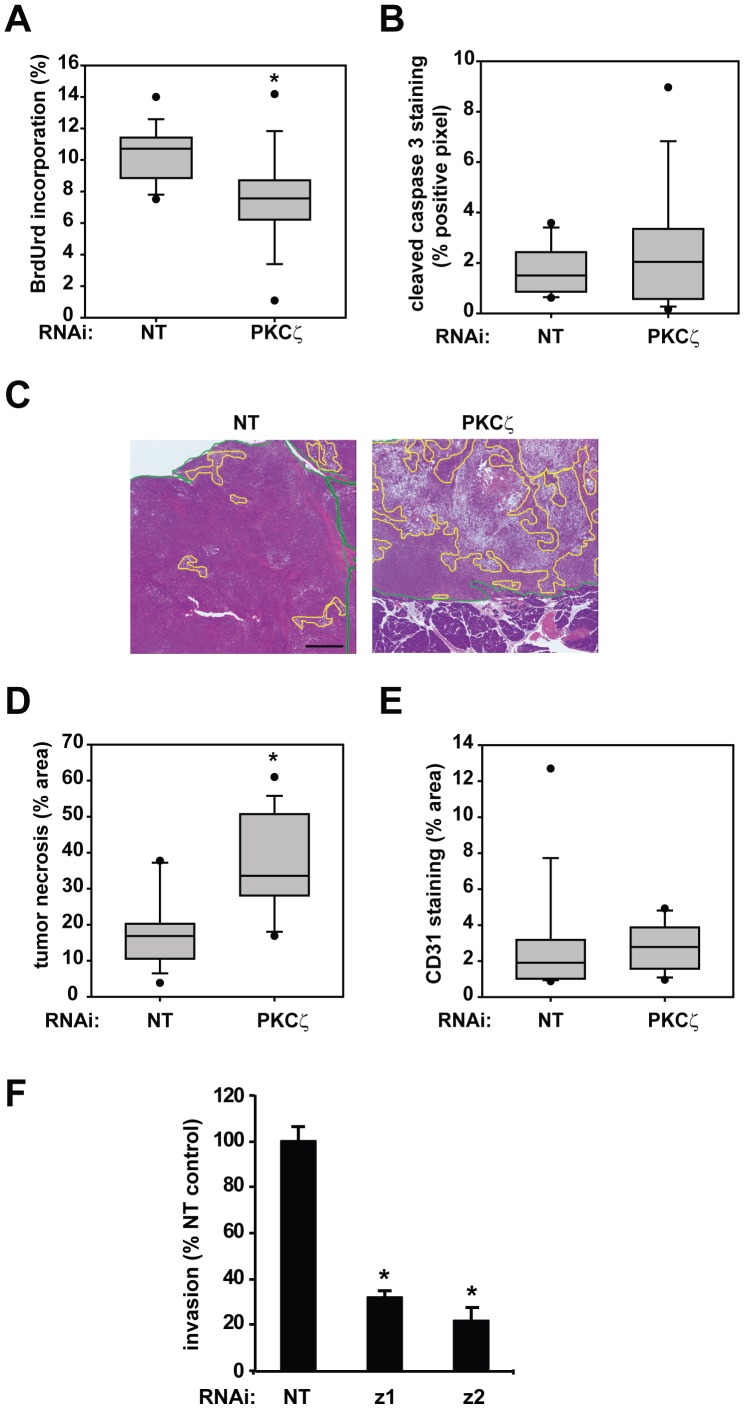
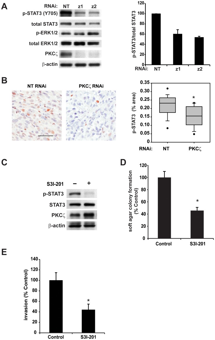
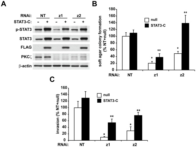
Pancreatic cancer is a very aggressive disease with few therapeutic options. In this study, we investigate the role of protein kinase C zeta (PKCζ) in pancreatic cancer cells. PKCζ has been shown to act as either a tumor suppressor or tumor promoter depending upon the cellular context. We find that PKCζ expression is either maintained or elevated in primary human pancreatic tumors, but is never lost, consistent with PKCζ playing a promotive role in the pancreatic cancer phenotype. Genetic inhibition of PKCζ reduced adherent growth, cell survival and anchorage-independent growth of human pancreatic cancer cells in vitro. Furthermore, PKCζ inhibition reduced orthotopic tumor size in vivo by inhibiting tumor cell proliferation and increasing tumor necrosis. In addition, PKCζ inhibition reduced tumor metastases in vivo, and caused a corresponding reduction in pancreatic cancer cell invasion in vitro. Signal transducer and activator of transcription 3 (STAT3) is often constitutively active in pancreatic cancer, and plays an important role in pancreatic cancer cell survival and metastasis. Interestingly, inhibition of PKCζ significantly reduced constitutive STAT3 activation in pancreatic cancer cells in vitro and in vivo. Pharmacologic inhibition of STAT3 mimicked the phenotype of PKCζ inhibition, and expression of a constitutively active STAT3 construct rescued the transformed phenotype in PKCζ-deficient cells. We conclude that PKCζ is required for pancreatic cancer cell transformed growth and invasion in vitro and tumorigenesis in vivo, and that STAT3 is an important downstream mediator of the pro-carcinogenic effects of PKCζ in pancreatic cancer cells.


Conflict of interest statement

Competing Interests: The authors have the following interests: A provisional patent related to this research has been filed (MSB, APF, NRM; Methods and Materials for treating pancreatic cancer, US Patent Application #20110190390). Co-author Alan Fields is an Academic Editor for PLOS ONE. There are no further patents, products in development or marketed products to declare. This does not alter the authors′ adherence to all the PLOS ONE policies on sharing data and materials.

Figures

Figure 1
Figure 1. PKCζ is elevated in a subset of human pancreatic tumors.
A and B) IHC detection of PKCζ expression in representative human pancreatic tumors (A; Tumor) and adjacent non-tumor tissue (B; Normal). B) Serial sections stained with H&E are provided to distinguish acinar, islet (top left image, pancreatic islet is outlined) and ductal cells (top right image). All images in the same panel are the same magnification. Bars = 100 µm. C) Quantitative PCR analysis of PKCζ mRNA expression was performed on 28 matched patient pancreatic adenocarcinoma and non-tumor samples. PKCζ expression was normalized to 18S abundance; *p = 0.001 calculated by paired t-test. D) PKCζ expression is significantly elevated in a subset of pancreatic tumors. PKCζ was overexpressed in 25% of pancreatic tumors analyzed, as defined by tumor mRNA abundance greater than 2 standard deviations above the average of PKCζ mRNA abundance in all adjacent non-tumor pancreas samples.
Figure 2
Figure 2. Inhibition of PKCζ expression reduces survival and transformed growth of pancreatic cancer cells.
Panc-1 cells stably carrying lentivirus expressing either control, non-targeting (NT) or PKCζ-targeting RNAi (z1 and z2) were assessed for A) PKCζ and PKCι protein expression by immunoblot analysis (top), and PKCζ mRNA abundance by qPCR analysis (bottom); B) cell viability (MTT colorimetric assay); C) cellular death (detected by Cell Death Detection ELISA) and D) anchorage-independent growth (colony formation in soft agar). For each panel Bars = average of 3 or more replicates±SD and graph is representative of 3 or more independent experiments. *p<0.05 vs NT.
Figure 3
Figure 3. Inhibition of PKCζ expression significantly reduces orthotopic pancreatic tumor size.
A) Representative bioluminescent imaging of mice with orthotopic Panc-1 NT and PKCζ RNAi pancreatic tumors. B) Representative H&E stained sections of orthotopic Panc-1 NT and PKCζ RNAi pancreatic tumors. The remaining normal mouse pancreas is circled in blue. C) Inhibition of PKCζ significantly decreased the pancreas and orthotopic tumor weight; n = 16; *p<0.001.
Figure 4
Figure 4. Inhibition of PKCζ expression significantly reduces orthotopic pancreatic tumor proliferation and increases tumor necrosis.
A) Quantitative analysis of tumor proliferation detected by BrdUrd incorporation; *p<0.003. B) Quantitative analysis of tumor apoptosis detected by cleaved caspase-3 staining. C) Representative H&E stained orthotopic Panc-1 NT and PKCζ RNAi pancreatic tumors with areas of necrosis identified (yellow outline) (bar = 1 mm). Green line delineates tumor tissue. D) Quantitative analysis of tumor necrosis plotted as percent of total tumor area; *p<0.0002. E) Quantitative analysis of tumor vascularity, as determined by percent area CD31 staining. A–E) n = 16 NT RNAi tumors and 15 PKCζ RNAi tumors. F) Panc-1 NT and PKCζ RNAi cells (z1 and z2) were assessed for cellular invasion through Matrigel-coated chambers. Bars = average of 3 or more replicates+/−SD and graph is representative of 2 or more independent experiments. *p<0.05 vs NT.
Figure 5
Figure 5. PKCζ expression regulates STAT3 phosphorylation.
A) Inhibition of PKCζ expression decreases constitutive STAT3 activation (detected as phospho-STAT3 Y705) but not ERK1/2 activation (detected as phospho-ERK1/2). Immunoblot analysis was performed on total cell lysates from Panc-1 NT and PKCζ RNAi cells (left) and expression analysis of immunoblot detection was performed (right) n = 3. B) Representative IHC detection of p-STAT3 in orthotopic Panc-1 NT and PKCζ RNAi pancreatic tumors (left), bar = 50 µm. Quantitative analysis of pSTAT3 IHC staining (right). C–E) The effect of STAT3 inhibitor (S3I-201) on C) STAT3 phosphorylation, D) anchorage-independent growth in soft agar and E) cellular invasion through Matrigel-coated chambers. In all assays, S3I-201 was used at 100 µm and an equal volume DMSO used as control diluent. For invasion assay, cells were pre-treated with S3I-201 or DMSO for 48 hours prior to initiation of the assay. Bars = average of 3 or more replicates+/−SD, and graph is representative of 2 or more independent experiments. *p<0.05.
Figure 6
Figure 6. Constitutively active STAT3 rescues the transformed phenotype in PKCζ RNAi-expressing cells.
Panc-1 cells expressing NT or PKCζ RNAi were infected with adenoviral constructs expressing either null (control), or constitutively active, FLAG-tagged STAT3 (STAT3-C). A) Immunoblot analysis of p-STAT3, STAT3, FLAG, PKCζ and β-actin expression. Cells were assessed for B) anchorage-independent growth in soft agar and C) cellular invasion through Matrigel-coated chambers. For each graph: Bars = average of 3 or more replicates±SD and graph is representative of 2 or more independent experiments. *significantly reduced compared to NT/null, p<0.05; **significantly increased compared to null-treated, p<0.05.

References

    1. Siegel R, Naishadham D, Jemal A (2012) Cancer statistics, 2012. CA Cancer J Clin 62: 10–29. - PubMed
    1. Lebedeva IV, Sarkar D, Su ZZ, Gopalkrishnan RV, Athar M, et al. (2006) Molecular target-based therapy of pancreatic cancer. Cancer Res 66: 2403–2413. - PubMed
    1. Castagna M, Takai Y, Kaibuchi K, Sano K, Kikkawa U, et al. (1982) Direct activation of calcium-activated, phospholipid-dependent protein kinase by tumor-promoting phorbol esters. J Biol Chem 257: 7847–7851. - PubMed
    1. Galvez AS, Duran A, Linares JF, Pathrose P, Castilla EA, et al. (2009) Protein kinase Cζ represses the interleukin-6 promoter and impairs tumorigenesis in vivo. Molecular and Cellular Biology 29: 104–115. - PMC - PubMed
    1. Murray NR, Jamieson L, Yu W, Zhang J, Gokmen-Polar Y, et al. (2004) Protein kinase C{iota} is required for Ras transformation and colon carcinogenesis in vivo. J Cell Biol 164: 797–802. - PMC - PubMed

Publication types

MeSH terms