. 2013 Aug 28;8(8):e72543.
doi: 10.1371/journal.pone.0072543. eCollection 2013.

Clioquinol synergistically augments rescue by zinc supplementation in a mouse model of acrodermatitis enteropathica

Affiliations

Clioquinol synergistically augments rescue by zinc supplementation in a mouse model of acrodermatitis enteropathica

Jim Geiser et al. PLoS One. .

Abstract

Background: Zinc deficiency due to poor nutrition or genetic mutations in zinc transporters is a global health problem and approaches to providing effective dietary zinc supplementation while avoiding potential toxic side effects are needed.

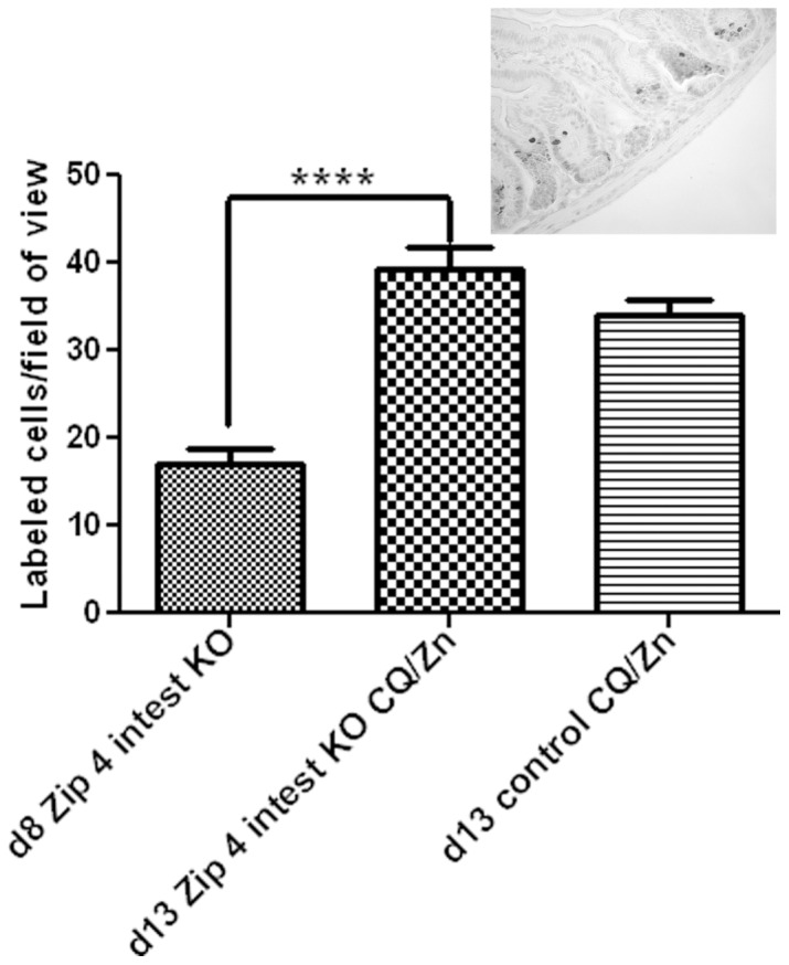
Methods/principal findings: Conditional knockout of the intestinal zinc transporter Zip4 (Slc39a4) in mice creates a model of the lethal human genetic disease acrodermatitis enteropathica (AE). This knockout leads to acute zinc deficiency resulting in rapid weight loss, disrupted intestine integrity and eventually lethality, and therefore provides a model system in which to examine novel approaches to zinc supplementation. We examined the efficacy of dietary clioquinol (CQ), a well characterized zinc chelator/ionophore, in rescuing the Zip4 (intest KO) phenotype. By 8 days after initiation of the knockout neither dietary CQ nor zinc supplementation in the drinking water was found to be effective at improving this phenotype. In contrast, dietary CQ in conjunction with zinc supplementation was highly effective. Dietary CQ with zinc supplementation rapidly restored intestine stem cell division and differentiation of secretory and the absorptive cells. These changes were accompanied by rapid growth and dramatically increased longevity in the majority of mice, as well as the apparent restoration of the homeostasis of several essential metals in the liver.

Conclusions: These studies suggest that oral CQ (or other 8-hydroxyquinolines) coupled with zinc supplementation could provide a facile approach toward treating zinc deficiency in humans by stimulating stem cell proliferation and differentiation of intestinal epithelial cells.


Conflict of interest statement

Competing Interests: Please note that co-author Paul A. Adlard is a PLOS ONE Editorial Board member, but this does not alter the authors' adherence to all the PLOS ONE policies on sharing data and materials.

Figures

Figure 1
Figure 1. Structure of clioquinol.
CQ is considered an intermediate affinity zinc chelator (Log conditional stability constant = 8.8; [67]) that also functions as a zinc ionophore. The ligand to metal stoichiometry is 1∶2 , . Its mechanism of action and clinical uses have been recently reviewed , .
Figure 2
Figure 2. Dietary CQ with excess zinc in the drinking water (CQ/Zn) can rescue Zip4 intest KO mice from the lethal effects of extreme zinc deficiency.
Mice homozygous for the floxed Zip4 gene and positive for the vil-CreERT2 gene (Zip4 intest KO) and littermates homozygous for the floxed Zip4 gene but negative for the vil-CreERT2 gene (control) were injected for 3 consecutive days with tamoxifen beginning 2 to 3 weeks after weaning and their body weights were monitored thereafter. These mice were fed normal chow and provided access to deionized water after weaning and then fed chow containing CQ and/or provided water containing excess zinc beginning on the indicated days. (A) Mice were fed chow containing CQ (CQ) or CQ chow plus water containing excess zinc (CQ/Zn) beginning 3 days after initiation of the intestine knockout. Only representative mice are shown for the sake of clarity in the figure. (B) On d8 after initiation of the intestine knockout mice were provided with water containing excess zinc. (C) On d8 after initiation of the intestine knockout these mice were fed chow containing CQ and provided access to drinking water containing excess zinc. Among these mice 6/8 thrived while (D) 2 did not thrive and one appeared to be blind in one eye.
Figure 3
Figure 3. Zip4 intest KO mice rescued by CQ/Zn remain dependent on CQ/Zn to survive.
Zip4 intest KO and control mice (n = 9 per group), as described in the legend to Figure 2, were fed normal chow and provided access to deionized water after weaning. On d8 after initiation of the knockout, mice were fed chow containing CQ and provided with water containing zinc (+CQ/Zn). On d22 the CQ/Zn diet was replaced with normal chow and deionized water (−CQ/Zn) and the mice were monitored thereafter. (A) Northern blot analysis of intestinal Zip4 mRNA from individual control mice (Con) and Zip4 intest KO mice on d8, d13 and d20 (4 mice per group; one sample is shown for d8, three are shown for d13 and two are shown for d20). (B) Body weights of Zip4 intest KO mice. (C) Body weights of control mice.
Figure 4
Figure 4. Quantification of phosphorylated-histone H3 labeled cells in sections of small intestine from Zip4 intest KO mice before and after feeding CQ/Zn. Zip4 intest KO mice and control mice (3 mice per group) were harvested on the indicated day after initiation of the knockout.
Mice were fed chow containing CQ and provided with water containing zinc (CQ/Zn) on d8 and harvested on d13. The small intestines were fixed, paraffin embedded and sections were processed for IHC using an antibody against Ser 10 phosphorylated-histone H3. The number of positive cells per field of view was counted in multiple fields of view (n = 14) at 400X magnification in several sections from 3 mice and the values shown are the mean ± S.E.M. Statistical significance was determined using the Unpaired T-test (two-tailed). ****indicates P<0.0001. Sections from knockout mice fed CQ/Zn that had no detectable Zip4 mRNA in the intestine were examined. Insert: A representative photomicrograph of the small intestine stained with antibody against phosphorylated-histone H3.
Figure 5
Figure 5. Development of the secretory and absorptive cell lineages in the small intestine is affected in Zip4 intest KO mice.
The small intestines from Zip4 intest KO mice were harvested on d8 after initiation of the knockout. (A) Paraffin sections were processed for IHC using antibodies against antigens specific to the secretory cell lineages (PC: Paneth cell lysozyme, GC: Goblet cell mucin 2 and EEC: Enteroendocrine cell chromogranin A). Brown color indicates positive immunostaining and the black arrows demarcate these cell types. (B) Left hand panels are lower magnification views of sections shown in (A). Panels on the right are paraffin sections stained with hematoxylin and eosin (top right), processed for IHC using an antibody against mouse fatty acid binding protein (Fabp2) which is enterocyte-specific (middle right) or antibody against mouse Sox9 which is expressed in stem cells and Paneth cells (lower right). These results were reproduced in multiple sections from 4 Zip4 intest KO mice (sections from 2 mice are shown), none of which had detectable Zip4 mRNA in the intestine.
Figure 6
Figure 6. Proper development of the secretory and absorptive cell lineages in the small intestine is rapidly restored in Zip4 intest KO mice fed CQ/Zn.
The small intestines from Zip4 intest KO mice were harvested on d13 after initiation of the knockout and starting CQ/Zn on d8. Sections from these mice which did not have detectable Zip4 mRNA were examined. (A: left hand panels) Paraffin sections were processed for IHC using antibodies against antigens specific to the secretory cell lineages (PC: Paneth cell lysozyme; GC: Goblet cell mucin 2; and EEC: Enteroendocrine cell chromogranin A). Brown color indicates positive immunostaining. (A: Right hand panels) Paraffin sections were stained with hematoxylin and eosin (top right), processed for IHC using an antibody against mouse fatty acid binding protein (Fabp2) which is enterocyte specific (middle right) or antibody against mouse Sox9 which is expressed in stem cells and Paneth cells (lower right). These results were reproduced in multiple sections from 4 Zip4 intest KO mice and the results were indistinguishable from those obtained using sections from control mice treated similarly. (B) Northern blot detection of Zip5 mRNA in the intestine from Zip4 intest KO mice on d8 and or d13 fed CQ/Zn. ZIP5 is a basolateral zinc transporter that is most abundant in the crypts but is also detectable on villus enterocytes .
Figure 7
Figure 7. ICP-MS quantification of essential metals in Zip4 intest KO mice before and after feeding CQ/Zn.
The small intestine, liver and pancreas from Zip4 intest KO mice were harvested on d8 after initiation of the knockout and on d13 and d20 after starting CQ/Zn on d8 (3 to 5 mice per group). Only data that showed significant changes are presented. Concentrations (ppm) for liver zinc (A), iron (B), copper (C), manganese (D), and magnesium (E), and intestine iron (F), were normalize to sulfur concentrations (ppm). Individual tissue samples were analyzed by ICP-MS for multiple elements including these essential metals. Data are expressed as metal/sulfur ratio ± S.E.M. Statistical significance was determined using the Unpaired T-test (two-tailed). Values were considered different if P<0.05. *indicates P<0.05; **indicates P<0.01; ***indicates P<0.001. Our previous studies showed that iron, copper and manganese accumulate in the liver when Zip4 is knocked out in the intestine whereas iron is reduced in the intestine . The symbol (>) on the Y-axes indicates approximate metal/sulfur ratios found in control mice.

References

    1. Prasad AS (2012) Discovery of human zinc deficiency: 50 years later. J Trace Elem Med Biol 26: 66–69. - PubMed
    1. Neggers YH, Cutter GR, Acton RT, Alvarez JO, Bonner JL et al (1990) A positive association between maternal serum zinc concentration and birth weight. Am J Clin Nutr 51: 678–684. - PubMed
    1. Tamura T, Goldenberg RL, Johnston KE, DuBard M (2000) Maternal plasma zinc concentrations and pregnancy outcome. Am J Clin Nutr 71: 109–113. - PubMed
    1. Hambidge KM, Krebs NF (2003) Zinc, low birth weight, and breastfeeding. Pediatrics 112: 1419–1420. - PubMed
    1. Bloxam DL, Bax CMR (1996) Zinc deficiency and abnormal fetal development: Assessment of maternal or fetal zinc status. Am J Obstet Gynecol 175: 1078. - PubMed

Publication types

MeSH terms

Supplementary concepts