. 2013 Sep 9:13:190.
doi: 10.1186/1471-2148-13-190.

Endosymbiosis in trypanosomatids: the genomic cooperation between bacterium and host in the synthesis of essential amino acids is heavily influenced by multiple horizontal gene transfers

Affiliations

Endosymbiosis in trypanosomatids: the genomic cooperation between bacterium and host in the synthesis of essential amino acids is heavily influenced by multiple horizontal gene transfers

João M P Alves et al. BMC Evol Biol. .

Abstract

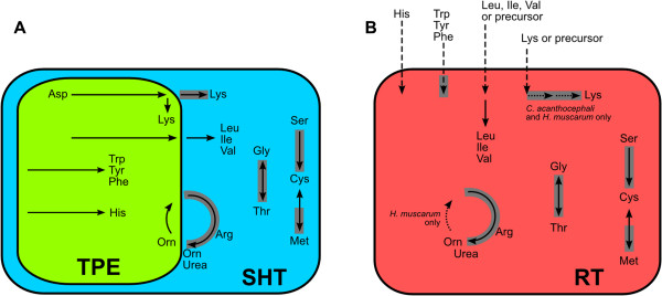
Background: Trypanosomatids of the genera Angomonas and Strigomonas live in a mutualistic association characterized by extensive metabolic cooperation with obligate endosymbiotic Betaproteobacteria. However, the role played by the symbiont has been more guessed by indirect means than evidenced. Symbiont-harboring trypanosomatids, in contrast to their counterparts lacking symbionts, exhibit lower nutritional requirements and are autotrophic for essential amino acids. To evidence the symbiont's contributions to this autotrophy, entire genomes of symbionts and trypanosomatids with and without symbionts were sequenced here.

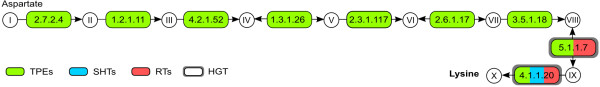
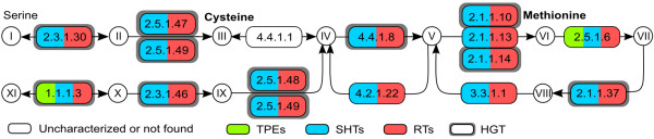
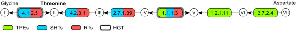
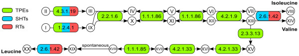
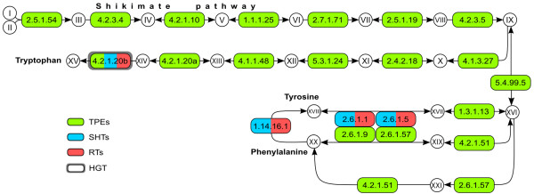
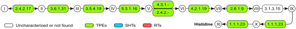
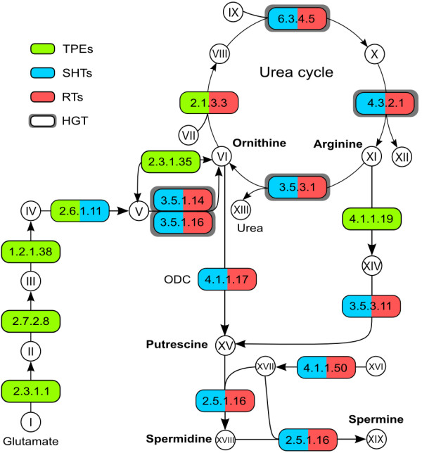
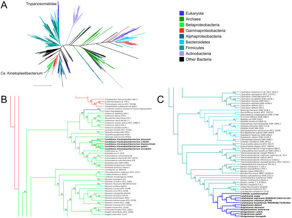
Results: Analyses of the essential amino acid pathways revealed that most biosynthetic routes are in the symbiont genome. By contrast, the host trypanosomatid genome contains fewer genes, about half of which originated from different bacterial groups, perhaps only one of which (ornithine cyclodeaminase, EC:4.3.1.12) derived from the symbiont. Nutritional, enzymatic, and genomic data were jointly analyzed to construct an integrated view of essential amino acid metabolism in symbiont-harboring trypanosomatids. This comprehensive analysis showed perfect concordance among all these data, and revealed that the symbiont contains genes for enzymes that complete essential biosynthetic routes for the host amino acid production, thus explaining the low requirement for these elements in symbiont-harboring trypanosomatids. Phylogenetic analyses show that the cooperation between symbionts and their hosts is complemented by multiple horizontal gene transfers, from bacterial lineages to trypanosomatids, that occurred several times in the course of their evolution. Transfers occur preferentially in parts of the pathways that are missing from other eukaryotes.

Conclusion: We have herein uncovered the genetic and evolutionary bases of essential amino acid biosynthesis in several trypanosomatids with and without endosymbionts, explaining and complementing decades of experimental results. We uncovered the remarkable plasticity in essential amino acid biosynthesis pathway evolution in these protozoans, demonstrating heavy influence of horizontal gene transfer events, from Bacteria to trypanosomatid nuclei, in the evolution of these pathways.


Figures

Figure 1
Figure 1
DAP pathway for lysine biosynthesis. Enzymes surrounded by a thick gray box were shown to be horizontally transferred from Bacteria (see main text). Metabolites – I: L-aspartate; II: 4-aspartyl-phosphate; III: aspartate 4-semialdehyde; IV: 2,3-dihydrodipicolinate; V: 2,3,4,5-tetrahydrodipicolinate; VI: N-succinyl-L-2-amino-6-oxopimelate; VII: N-succinyl-LL-2,6-diaminopimelate; VIII: LL-2,6-diaminopimelate; IX: meso-2,6-diaminopimelate; X: lysine. Enzymes – 2.7.2.4: aspartate kinase; 1.2.1.11: aspartate-semialdehyde dehydrogenase; 4.2.1.52: dihydrodipicolinate synthase; 1.3.1.26: dihydrodipicolinate reductase; 2.3.1.117: tetrahydrodipicolinate succinyltransferase; 2.6.1.17: succinyldiaminopimelate transaminase; 3.5.1.18: succinyldiaminopimelate desuccinylase; 5.1.1.7: diaminopimelate epimerase; 4.1.1.20: diaminopimelate decarboxylase. SHT: symbiont-harboring trypanosomatid; RT: regular trypanosomatid; TPE: trypanosomatid proteobacterial endosymbiont.
Figure 2
Figure 2
Cysteine and methionine synthesis and interconversion pathway. Enzymes surrounded by a thick gray box were shown to be horizontally transferred from Bacteria (see main text). Metabolites – I: L-serine; II: O-acetyl-serine; III: cysteine; IV: cystathionine; V: homocysteine; VI: methionine; VII: S-adenosyl-methionine; VIII: S-adenosyl-homocysteine; IX: succinyl-homoserine; X: homoserine; XI: aspartate 4-semialdehyde. Enzymes – 2.3.1.30: serine O-acetyltransferase; 2.5.1.47: cysteine synthase; 4.4.1.1: cystathionine gamma-lyase; 4.4.1.8: cystathionine beta-lyase; 2.1.1.x: 2.1.1.10, homocysteine S-methyltransferase, 2.1.1.13, 5-methyltetrahydrofolate–homocysteine methyltransferase, 2.1.1.14, 5-methyltetrahydropteroyltriglutamate–homocysteine methyltransferase; 2.5.1.6: S-adenosyl-methionine synthetase; 2.1.1.37: DNA (cytosine-5-)-methyltransferase; 3.3.1.1: adenosylhomocysteinase; 4.2.1.22: cystathionine beta-synthase; 2.5.1.48: cystathionine gamma-synthase; 2.3.1.46: homoserine O-succinyltransferase; 1.1.1.3: homoserine dehydrogenase. Abbreviations as for Figure 1.
Figure 3
Figure 3
Threonine synthesis pathway. Enzymes surrounded by a thick gray box were shown to be horizontally transferred from Bacteria (see main text). Metabolites – I: glycine; II: threonine; III: phosphohomoserine; IV: homoserine; V: aspartate 4-semialdehyde; VI: 4-aspartyl-phosphate; VII: L-aspartate. Enzymes – 4.1.2.5: threonine aldolase; 2.7.2.4: aspartate kinase; 1.2.1.11: aspartate-semialdehyde dehydrogenase; 1.1.1.3: homoserine dehydrogenase; 2.7.1.39: homoserine kinase; 4.2.3.1: threonine synthase. Abbreviations as for Figure 1.
Figure 4
Figure 4
Isoleucine, valine, and leucine synthesis pathway. Metabolites – I: pyruvate; II: threonine; III: 2-oxobutanoate; IV: (S)-2-aceto-2-hydroxybutanoate; V: (R)-3-hydroxy-3-methyl-2-oxopentanoate; VI: (R)-2,3-dihydroxy-3-methylpentanoate; VII: (S)-3-methyl-2-oxopentanoate; VIII: isoleucine; IX: 2-(alpha-hydroxyethyl) thiamine diphosphate; X: (S)-2-acetolactate; XI: 3-hydroxy-3-methyl-2-oxobutanoate; XII: (R)-2,3-dihydroxy-3-methylbutanoate; XIII: 2-oxoisovalerate; XIV: valine; XV: (2S)-2-isopropylmalate; XVI: 2-isopropylmaleate; XVII: (2R,3S)-3-isopropylmalate; XVIII: (2S)-2-isopropyl-3-oxosuccinate; XIX: 4-methyl-2-oxopentanoate; XX: leucine. Enzymes – 1.2.4.1: pyruvate dehydrogenase E1 component subunit alpha; 4.3.1.19: threonine ammonia-lyase; 2.2.1.6: acetolactate synthase small and large subunits; 1.1.1.86: ketol-acid reductoisomerase; 4.2.1.9: dihydroxy-acid dehydratase; 2.6.1.42: branched-chain amino acid transaminase; 2.3.3.13: 2-isopropylmalate synthase; 4.2.1.33: 3-isopropylmalate dehydratase small and large subunits; 1.1.1.85: 3-isopropylmalate dehydrogenase. Abbreviations as for Figure 1.
Figure 5
Figure 5
Phenylalanine, tyrosine, and tryptophan synthesis pathway. Enzymes surrounded by a thick gray box were shown to be horizontally transferred from Bacteria (see main text). Metabolites – I: D-erythrose 4-phosphate; II: phosphoenolpyruvate; III: 7-phosphate-2-dehydro-3-deoxy-D-arabinoheptonate; IV: 3-dehydroquinate; V: 3-dehydroshikimate; VI: shikimate; VII: shikimate 3-phosphate; VIII: 5-O-(1-carboxyvinyl)-3-phosphoshikimate; IX: chorismate; X: anthranilate; XI: N-(5-Phospho-D-ribosyl)anthranilate; XII: 1-(2-carboxyphenylamino)-1-deoxy-D-ribulose 5-phosphate; XIII: indoleglycerol phosphate; XIV: indole; XV: tryptophan; XVI: prephenate; XVII: arogenate; XVIII: phenylpyruvate; XIX: 4-hydroxyphenylpyruvate; XX: tyrosine; XXI: phenylalanine. Enzymes – 2.5.1.54: 3-deoxy-7-phosphoheptulonate synthase; 4.2.3.4: 3-dehydroquinate synthase; 4.2.1.10: 3-dehydroquinate dehydratase I; 1.1.1.25: shikimate dehydrogenase; 2.7.1.71: shikimate kinase; 2.5.1.19: 3-phosphoshikimate 1-carboxyvinyltransferase; 4.2.3.5: chorismate synthase; 4.1.3.27: anthranilate synthase; 2.4.2.18: anthranilate phosphoribosyltransferase; 5.3.1.24: phosphoribosylanthranilate isomerase; 4.1.1.48: indoleglycerol phosphate synthetase; 4.2.1.20a/b: tryptophan synthase alpha (a) and beta (b) subunits; 4.2.1.51/5.4.99.5: bifunctional prephenate dehydratase/chorismate mutase; 2.6.1.57: aromatic amino acid aminotransferase; 1.3.1.13: prephenate dehydrogenase (NADP+); 2.6.1.1: aspartate aminotransferase; 2.6.1.5: tyrosine aminotransferase; 2.6.1.9: histidinol-phosphate aminotransferase; 1.14.16.1: phenylalanine-4-hydroxylase. Abbreviations as for Figure 1.
Figure 6
Figure 6
Histidine synthesis pathway. Enzymes surrounded by a thick gray box were shown to be horizontally transferred from Bacteria (see main text). Metabolites – I: 5-phosphoribosyl diphosphate (PRPP); II: phosphoribosyl-ATP; III: phosphoribosyl-AMP; IV: phosphoribosyl-formimino-AICAR phosphate; V: phosphoribulosyl-formimino-AICAR phosphate; VI: imidazole-glycerol 3-phosphate; VII: imidazole-acetol phosphate; VIII: histidinol phosphate; IX: histidinol; X: histidinal; XI: histidine. Enzymes – 2.4.2.17: ATP phosphoribosyltransferase; 3.6.1.31: phosphoribosyl-ATP pyrophosphohydrolase; 3.5.4.19: phosphoribosyl-AMP cyclohydrolase; 5.3.1.16: phosphoribosylformimino-5-aminoimidazole carboxamide ribotide isomerase; 4.1.3.-: cyclase HisF; 2.4.2.-: glutamine amidotransferase; 4.2.1.19: imidazole-glycerol phosphate dehydratase; 2.6.1.9: histidinol phosphate aminotransferase; 3.1.3.15: histidinol phosphatase; 1.1.1.23: histidinol dehydrogenase. Abbreviations as for Figure 1.
Figure 7
Figure 7
Arginine, ornithine, and polyamine synthesis pathway. Enzymes surrounded by a thick gray box were shown to be horizontally transferred from Bacteria (see main text). Metabolites – I: glutamate; II: N-acetylglutamate; III: N-acetylglutamyl-phosphate; IV: N-acetyl-glutamate semialdehyde; V: N-acetylornithine; VI: ornithine; VII: carbamoyl-phosphate; VIII: citruline; IX: aspartate; X: arginino succinate; XI: arginine; XII: fumarate; XIII: urea; XIV: agmatine; XV: putrescine; XVI: S-adenosylmethionine; XVII: S-adenosylmethioninamine; XVIII: spermidine; XIX: spermine. Enzymes – 2.1.3.3: ornithine carbamoyltransferase; 6.3.4.5: argininosuccinate synthase; 4.3.2.1: argininosuccinate lyase; 3.5.3.1: arginase; 4.1.1.17: ornithine decarboxylase; 3.5.1.14: aminoacylase; 3.5.1.16: acetylornithine deacetylase; 2.3.1.35: glutamate N-acetyltransferase; 2.6.1.11: acetylornithine aminotransferase; 1.2.1.38: N-acetyl-gamma-glutamyl-phosphate reductase; 2.7.2.8: acetylglutamate kinase; 2.3.1.1: amino-acid N-acetyltransferase; 3.5.3.11: agmatinase; 4.1.1.19: arginine decarboxylase; 4.1.1.50: adenosylmethionine decarboxylase; 2.5.1.16: spermidine synthase. Abbreviations as for Figure 1.
Figure 8
Figure 8
Maximum likelihood phylogenetic tree of homoserine dehydrogenase (EC:1.1.1.3). A – overall tree, colored according to taxonomic affiliation of each taxon, as per the legend on the right; distance bar only applies to panel A. B – details of the region of the tree where the Ca. Kinetoplastibacterium spp. are placed. C – details of the region of the tree where the Trypanosomatidae are placed. Values on nodes represent bootstrap support (only 50 or greater shown). Panels B and C are meant to only represent the branching patterns and do not portray estimated distances between sequences. Abbreviations as for Figure 1.
Figure 9
Figure 9
Overview of the biosynthetic pathways of essential amino acids in trypanosomatids. Dashed arrows: metabolite import; dotted arrows: reaction present in only some of the organisms analyzed; solid arrows: other reactions (a single arrow can summarize multiple steps); arrows surrounded by a gray box: enzymes possibly acquired through horizontal transfer from Bacteria to trypanosomatids (see main text). A. Contribution of symbiont-harboring trypanosomatids (SHT) and endosymbionts (TPE) based on the analysis of gene content in the genomes of A. deanei, A. desouzai, S. culicis, S. oncopelti, S. galati, and respective endosymbionts. B. Biochemical capability of trypanosomatids without symbionts (RT), based on the analysis of genomic data from H. muscarum, C. acanthocephali, and L. major.

References

    1. Baumann P, Moran NA, Baumann L. The evolution and genetics of aphid endosymbionts. BioScience. 1997;47:12–20. doi: 10.2307/1313002. - DOI
    1. Wernegreen JJ. Genome evolution in bacterial endosymbionts of insects. Nat Rev Genet. 2002;3:850–861. doi: 10.1038/nrg931. - DOI - PubMed
    1. Wernegreen JJ. Endosymbiosis: lessons in conflict resolution. PLoS Biol. 2004;2:E68. doi: 10.1371/journal.pbio.0020068. - DOI - PMC - PubMed
    1. Moran NA. Symbiosis. Curr Biol. 2006;16:R866–R871. doi: 10.1016/j.cub.2006.09.019. - DOI - PubMed
    1. Moran NA, McCutcheon JP, Nakabachi A. Genomics and evolution of heritable bacterial symbionts. Annu Rev Genet. 2008;42:165–190. doi: 10.1146/annurev.genet.41.110306.130119. - DOI - PubMed

Publication types

Substances

LinkOut - more resources