Inhibition of cereal rust fungi by both class I and II defensins derived from the flowers of Nicotiana alata
- PMID: 24015961
- PMCID: PMC6638682
- DOI: 10.1111/mpp.12066
Inhibition of cereal rust fungi by both class I and II defensins derived from the flowers of Nicotiana alata
Abstract
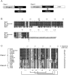
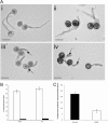
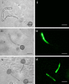
Defensins are a large family of small, cysteine-rich, basic proteins, produced by most plants and plant tissues. They have a primary function in defence against fungal disease, although other functions have been described. This study reports the isolation and characterization of a class I secreted defensin (NaD2) from the flowers of Nicotiana alata, and compares its antifungal activity with the class II defensin (NaD1) from N. alata flowers, which is stored in the vacuole. NaD2, like all other class I defensins, lacks the C-terminal pro-peptide (CTPP) characteristic of class II defensins. NaD2 is most closely related to Nt-thionin from N. tabacum (96% identical) and shares 81% identity with MtDef4 from alfalfa. The concentration required to inhibit in vitro fungal growth by 50% (IC50 ) was assessed for both NaD1 and NaD2 for the biotrophic basidiomycete fungi Puccinia coronata f. sp. avenae (Pca) and P. sorghi (Ps), the necrotrophic pathogenic ascomycetes Fusarium oxysporum f. sp. vasinfectum (Fov), F. graminearum (Fgr), Verticillium dahliae (Vd) and Thielaviopsis basicola (Tb), and the saprobe Aspergillus nidulans. NaD1 was a more potent antifungal molecule than NaD2 against both the biotrophic and necrotrophic fungal pathogens tested. NaD2 was 5-10 times less effective at killing necrotrophs, but only two-fold less effective on Puccinia species. A new procedure for testing antifungal proteins is described in this study which is applicable to pathogens with spores that are not amenable to liquid culture, such as rust pathogens. Rusts are the most damaging fungal pathogens of many agronomically important crop species (wheat, barley, oats and soybean). NaD1 and NaD2 inhibited urediniospore germination, germ tube growth and germ tube differentiation (appressoria induction) of both Puccinia species tested. NaD1 and NaD2 were fungicidal on Puccinia species and produced stunted germ tubes with a granular cytoplasm. When NaD1 and NaD2 were sprayed onto susceptible oat plants prior to the plants being inoculated with crown rust, they reduced the number of pustules per leaf area, as well as the amount of chlorosis induced by infection. Similar to observations in vitro, NaD1 was more effective as an antifungal control agent than NaD2. Further investigation revealed that both NaD1 and NaD2 permeabilized the plasma membranes of Puccinia spp. This study provides evidence that both secreted (NaD2) and nonsecreted (NaD1) defensins may be useful for broad-spectrum resistance to pathogens.
© 2013 BSPP AND JOHN WILEY & SONS LTD.
Figures


Similar articles
-
Plant Defensins NaD1 and NaD2 Induce Different Stress Response Pathways in Fungi.Int J Mol Sci. 2016 Sep 3;17(9):1473. doi: 10.3390/ijms17091473. Int J Mol Sci. 2016. PMID: 27598152 Free PMC article.
-
The C-terminal propeptide of a plant defensin confers cytoprotective and subcellular targeting functions.BMC Plant Biol. 2014 Feb 5;14:41. doi: 10.1186/1471-2229-14-41. BMC Plant Biol. 2014. PMID: 24495600 Free PMC article.
-
The plant defensin, NaD1, enters the cytoplasm of Fusarium oxysporum hyphae.J Biol Chem. 2008 May 23;283(21):14445-52. doi: 10.1074/jbc.M709867200. Epub 2008 Mar 13. J Biol Chem. 2008. PMID: 18339623
-
Puccinia coronata f. sp. avenae: a threat to global oat production.Mol Plant Pathol. 2018 May;19(5):1047-1060. doi: 10.1111/mpp.12608. Epub 2017 Dec 10. Mol Plant Pathol. 2018. PMID: 28846186 Free PMC article. Review.
-
Role of Alternate Hosts in Epidemiology and Pathogen Variation of Cereal Rusts.Annu Rev Phytopathol. 2016 Aug 4;54:207-28. doi: 10.1146/annurev-phyto-080615-095851. Epub 2016 Jan 1. Annu Rev Phytopathol. 2016. PMID: 27296143 Review.
Cited by
-
Resistance to the Plant Defensin NaD1 Features Modifications to the Cell Wall and Osmo-Regulation Pathways of Yeast.Front Microbiol. 2018 Jul 24;9:1648. doi: 10.3389/fmicb.2018.01648. eCollection 2018. Front Microbiol. 2018. PMID: 30087664 Free PMC article.
-
Characterization and functional analysis of gerbera plant defensin (PDF) genes reveal the role of GhPDF2.4 in defense against the root rot pathogen Phytophthora cryptogea.aBIOTECH. 2024 Mar 31;5(3):325-338. doi: 10.1007/s42994-024-00146-8. eCollection 2024 Sep. aBIOTECH. 2024. PMID: 39279851 Free PMC article.
-
Ethnobotany and Antimicrobial Peptides From Plants of the Solanaceae Family: An Update and Future Prospects.Front Pharmacol. 2020 May 7;11:565. doi: 10.3389/fphar.2020.00565. eCollection 2020. Front Pharmacol. 2020. PMID: 32477108 Free PMC article. Review.
-
Overexpression of a defensin enhances resistance to a fruit-specific anthracnose fungus in pepper.PLoS One. 2014 May 21;9(5):e97936. doi: 10.1371/journal.pone.0097936. eCollection 2014. PLoS One. 2014. PMID: 24848280 Free PMC article.
-
Fighting pathogenic yeasts with plant defensins and anti-fungal proteins from fungi.Appl Microbiol Biotechnol. 2024 Mar 27;108(1):277. doi: 10.1007/s00253-024-13118-1. Appl Microbiol Biotechnol. 2024. PMID: 38536496 Free PMC article. Review.
References
-
- Anikster, Y. and Wahl, I. (1979) Coevolution of the rust fungi on Gramineae and Liliaceae and their hosts. Annu. Rev. Phytopathol. 17, 367–403.
-
- Ayliffe, M. , Singh, R. and Lagudah, E. (2008) Durable resistance to wheat stem rust needed. Curr. Opin. Plant Biol. 11, 187–192. - PubMed
-
- Barna, B. , Leiter, É. , Hegedus, N. , Bíró, T. and Pócsi, I. (2008) Effect of the Penicillium chrysogenum antifungal protein (PAF) on barley powdery mildew and wheat leaf rust pathogens. J. Basic Microbiol. 48, 516–520. - PubMed
-
- Broekaert, W. , Cammue, B. , De Bolle, M. , Thevissen, K. , De Samblanx, G. and Osborne, R. (1997) Antimicrobial peptides from plants. Crit. Rev. Plant Sci. 16, 297–323.
-
- Bulet, P. , Hetru, C. , Dimarcq, J. and Hoffmann, D. (1999) Antimicrobial peptides in insects; structure and function. Dev. Comp. Immunol. 23, 329–344. - PubMed
MeSH terms
Substances
LinkOut - more resources
Full Text Sources
Other Literature Sources
Research Materials
Miscellaneous

