

Selective nucleic acid removal via exclusion (SNARE): capturing mRNA and DNA from a single sample
- PMID: 24016179
- PMCID: PMC3897163
- DOI: 10.1021/ac402162r
Selective nucleic acid removal via exclusion (SNARE): capturing mRNA and DNA from a single sample
Abstract
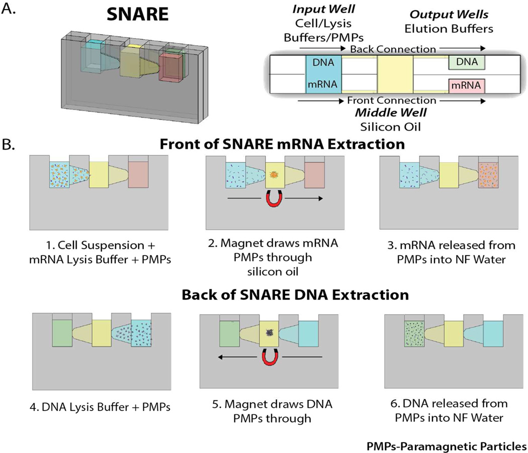
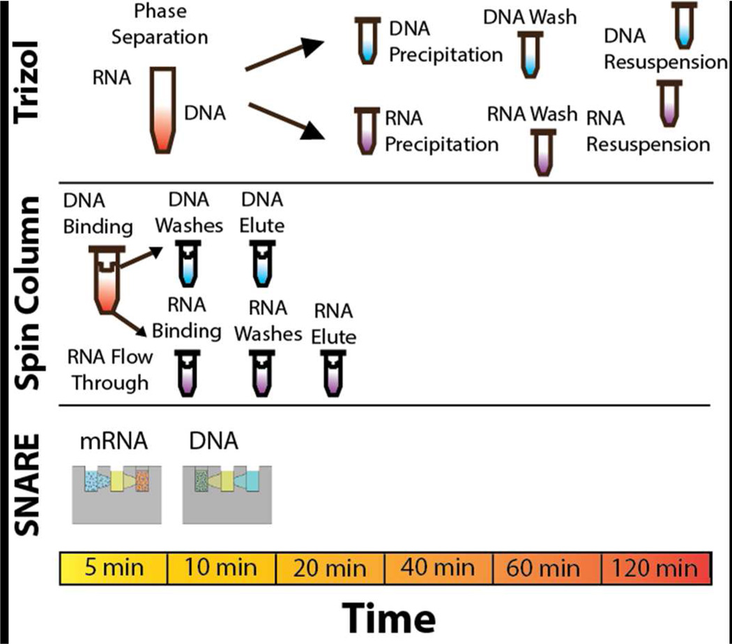
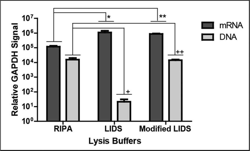
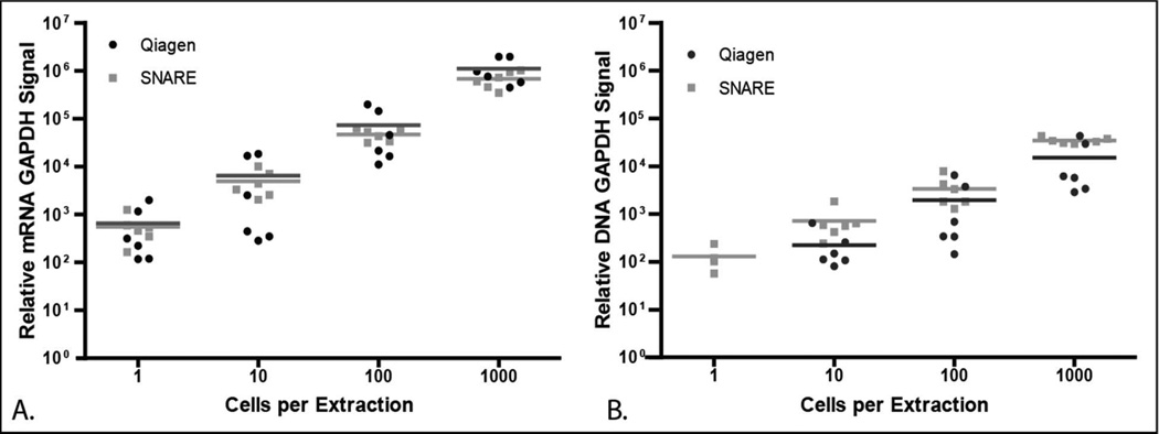
The path from gene (DNA) to gene product (RNA or protein) is the foundation of genotype giving rise to phenotype. Comparison of genomic analyses (DNA) with paired transcriptomic studies (mRNA) is critical to evaluating the pathogenic processes that give rise to human disease. The ability to analyze both DNA and mRNA from the same sample is not only important for biologic interrogation but also to minimize variance (e.g., sample loss) unrelated to the biology. Existing methods for RNA and DNA purification from a single sample are typically time-consuming and labor intensive or require large sample sizes to split for separate RNA and DNA extraction procedures. Thus, there is a need for more efficient and cost-effective methods to purify both RNA and DNA from a single sample. To address this need, we have developed a technique, termed SNARE (Selective Nucleic Acid Removal via Exclusion), that uses pinned oil interfaces to simultaneous purify mRNA and DNA from a single sample. A unique advantage of SNARE is the elimination of dilutive wash and centrifugation processes that are fundamental to conventional methods where sample is typically discarded. This minimizes loss and maximizes recovery by allowing nondilutive reinterrogation of the sample. We demonstrate that SNARE is more sensitive than commercially available kits, robustly and repeatably achieving mRNA and DNA purification from extremely low numbers of cells for downstream analyses. In addition to sensitivity, SNARE is fast, easy to use, and cost-effective and requires no laboratory infrastructure or hazardous chemicals. We demonstrate the clinical utility of the SNARE with prostate cancer circulating tumor cells to demonstrate its ability to perform both genomic and transcriptomic interrogation on rare cell populations that would be difficult to achieve with any current method.
Figures
References
-
- Cirilo PD, Marchi FA, Barros Filho Mde C, Rocha RM, Domingues MA, Jurisica I, Pontes A, Rogatto SR. PloS one. 2013;8:e57901. - PMC - PubMed
- Curtis C, Shah SP, Chin SF, Turashvili G, Rueda OM, Dunning MJ, Speed D, Lynch AG, Samarajiwa S, Yuan Y, Graf S, Ha G, Haffari G, Bashashati A, Russell R, McKinney S, Group M, Langerod A, Green A, Provenzano E, Wishart G, Pinder S, Watson P, Markowetz F, Murphy L, Ellis I, Purushotham A, Borresen-Dale AL, Brenton JD, Tavare S, Caldas C, Aparicio S. Nature. 2012;486:346–352. - PMC - PubMed
- Wu C, Wyatt AW, Lapuk AV, McPherson A, McConeghy BJ, Bell RH, Anderson S, Haegert A, Brahmbhatt S, Shukin R, Mo F, Li E, Fazli L, Hurtado-Coll A, Jones EC, Butterfield YS, Hach F, Hormozdiari F, Hajirasouliha I, Boutros PC, Bristow RG, Jones SJ, Hirst M, Marra MA, Maher CA, Chinnaiyan AM, Sahinalp SC, Gleave ME, Volik SV, Collins CC. The Journal of pathology. 2012;227:53–61. - PMC - PubMed
-
- Mathieson W, Thomas GA. Analytical biochemistry. 2013;433:10–18. - PubMed
-
- Grzendowski M, Riemenschneider MJ, Hawranke E, Stefanski A, Meyer HE, Reifenberger G, Stuhler K. Proteomics. 2009;9:4985–4990. - PubMed
-
- Chomczynski P. BioTechniques. 1993;15:532–534. 536–537. - PubMed
Publication types
MeSH terms
Substances
Grants and funding
LinkOut - more resources
Full Text Sources
Other Literature Sources
