

Expanding horizons: ciliary proteins reach beyond cilia
- PMID: 24016188
- PMCID: PMC5703194
- DOI: 10.1146/annurev-genet-111212-133243
Expanding horizons: ciliary proteins reach beyond cilia
Abstract
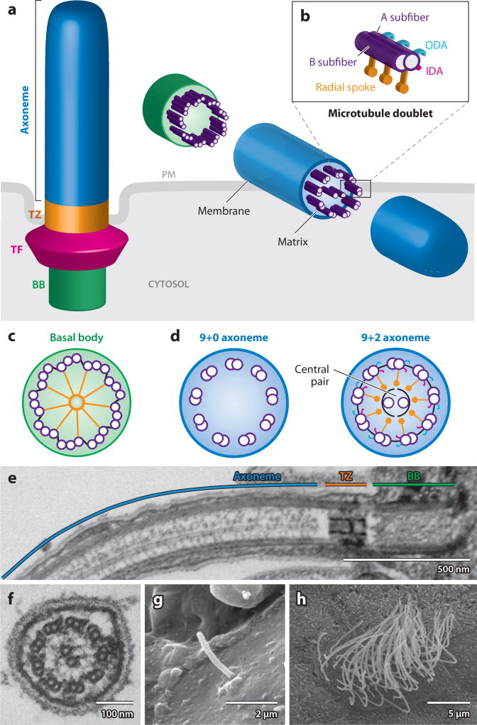
Once obscure, the cilium has come into the spotlight during the past decade. It is now clear that aside from generating locomotion by motile cilia, both motile and immotile cilia serve as signaling platforms for the cell. Through both motility and sensory functions, cilia play critical roles in development, homeostasis, and disease. To date, the cilium proteome contains more than 1,000 different proteins, and human genetics is identifying new ciliopathy genes at an increasing pace. Although assigning a function to immotile cilia was a challenge not so long ago, the myriad of signaling pathways, proteins, and biological processes associated with the cilium have now created a new obstacle: how to distill all these interactions into specific themes and mechanisms that may explain how the organelle serves to maintain organism homeostasis. Here, we review the basics of cilia biology, novel functions associated with cilia, and recent advances in cilia genetics, and on the basis of this framework, we further discuss the meaning and significance of ciliary connections.
Figures
Similar articles
-
Ciliopathies and the Kidney: A Review.Am J Kidney Dis. 2021 Mar;77(3):410-419. doi: 10.1053/j.ajkd.2020.08.012. Epub 2020 Oct 9. Am J Kidney Dis. 2021. PMID: 33039432 Review.
-
Active transport and diffusion barriers restrict Joubert Syndrome-associated ARL13B/ARL-13 to an Inv-like ciliary membrane subdomain.PLoS Genet. 2013;9(12):e1003977. doi: 10.1371/journal.pgen.1003977. Epub 2013 Dec 5. PLoS Genet. 2013. PMID: 24339792 Free PMC article.
-
Novel transglutaminase-like peptidase and C2 domains elucidate the structure, biogenesis and evolution of the ciliary compartment.Cell Cycle. 2012 Oct 15;11(20):3861-75. doi: 10.4161/cc.22068. Epub 2012 Sep 14. Cell Cycle. 2012. PMID: 22983010 Free PMC article.
-
New Insights into Cystic Kidney Diseases.Contrib Nephrol. 2018;195:31-41. doi: 10.1159/000486932. Epub 2018 May 7. Contrib Nephrol. 2018. PMID: 29734148 Review.
-
[Hereditary cerebro-oculo-renal syndromes].Ugeskr Laeger. 2014 Feb 17;176(8A):V07130452. Ugeskr Laeger. 2014. PMID: 25350305 Review. Danish.
Cited by
-
Structure of the N-terminal coiled-coil domains of the ciliary protein Rpgrip1l.iScience. 2023 Feb 21;26(3):106249. doi: 10.1016/j.isci.2023.106249. eCollection 2023 Mar 17. iScience. 2023. PMID: 36915689 Free PMC article.
-
Genetic contributors and modifiers of biliary atresia.Dig Dis. 2015;33(3):408-14. doi: 10.1159/000371694. Epub 2015 May 27. Dig Dis. 2015. PMID: 26045276 Free PMC article. Review.
-
NHERF1/EBP50 is an organizer of polarity structures and a diagnostic marker in ependymoma.Acta Neuropathol Commun. 2015 Mar 8;3:11. doi: 10.1186/s40478-015-0197-z. Acta Neuropathol Commun. 2015. PMID: 25775275 Free PMC article.
-
Dissecting the Vesicular Trafficking Function of IFT Subunits.Front Cell Dev Biol. 2020 Jan 15;7:352. doi: 10.3389/fcell.2019.00352. eCollection 2019. Front Cell Dev Biol. 2020. PMID: 32010685 Free PMC article. Review.
-
Deacetylation of α-tubulin and cortactin is required for HDAC6 to trigger ciliary disassembly.Sci Rep. 2015 Aug 6;5:12917. doi: 10.1038/srep12917. Sci Rep. 2015. PMID: 26246421 Free PMC article.
References
-
- Abe G, Suster ML, Kawakami K. Tol2-mediated transgenesis, gene trapping, enhancer trapping, and the Gal4-UAS system. Methods Cell Biol. 2011;104:23–49. - PubMed
-
- Afzelius BA. A human syndrome caused by immotile cilia. Science. 1976;193:317–19. - PubMed
-
- Anderson KV. Cilia and Hedgehog signaling in the mouse embryo. Harvey Lect. 2006;102:103–15. - PubMed
-
- Ansley SJ, Badano JL, Blacque OE, Hill J, Hoskins BE, et al. Basal body dysfunction is a likely cause of pleiotropic Bardet-Biedl syndrome. Nature. 2003;425:628–33. - PubMed
Publication types
MeSH terms
Substances
Supplementary concepts
Grants and funding
LinkOut - more resources
Full Text Sources
Other Literature Sources
Miscellaneous
