. 2013 Dec;34(37):9244-51.
doi: 10.1016/j.biomaterials.2013.08.042. Epub 2013 Sep 7.

The influence of substrate topography on the migration of corneal epithelial wound borders

Affiliations

The influence of substrate topography on the migration of corneal epithelial wound borders

Bernardo Yanez-Soto et al. Biomaterials. 2013 Dec.

Abstract

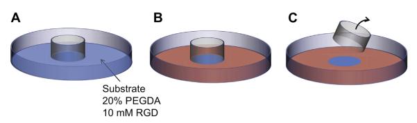
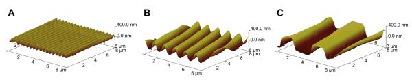
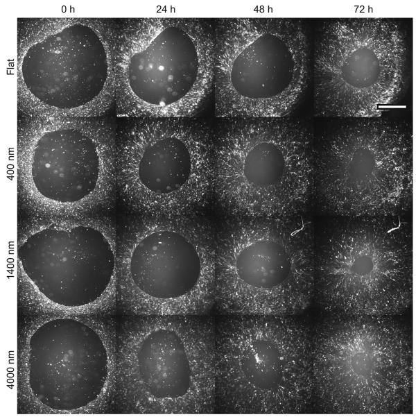
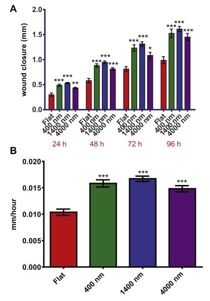
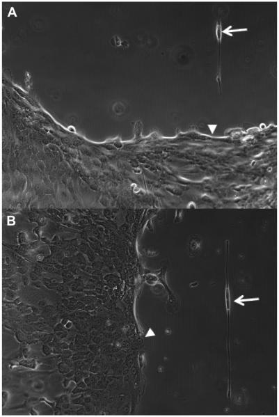
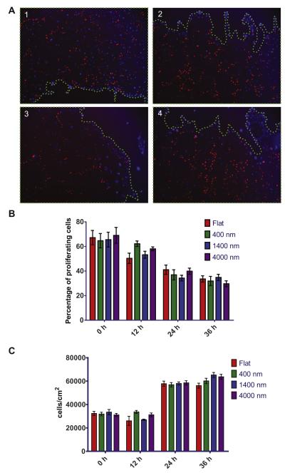
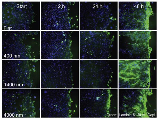
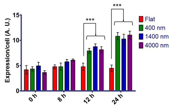
Currently available artificial corneas can develop post-implant complications including epithelial downgrowth, infection, and stromal melting. The likelihood of developing these disastrous complications could be minimized through improved formation and maintenance of a healthy epithelium covering the implant. We hypothesize that this epithelial formation may be enhanced through the incorporation of native corneal basement membrane biomimetic chemical and physical cues onto the surface of the keratoprosthesis. We fabricated hydrogel substrates molded with topographic features containing specific bio-ligands and developed an in vitro wound healing assay. In our experiments, the rate of corneal epithelial wound healing was significantly increased by 50% in hydrogel surfaces containing topographic features, compared to flat surfaces with the same chemical attributes. We determined that this increased healing is not due to enhanced proliferation or increased spreading of the epithelial cells, but to an increased active migration of the epithelial cells. These results show the potential benefit of restructuring and improving the surface of artificial corneas to enhance epithelial coverage and more rapidly restore the formation of a functional epithelium.

Keywords: Biomimetic material; Corneal wound healing; Epithelial cell; Hydrogel; Nanotopography; Polyethylene glycol.


Figures

Fig. 1
Fig. 1
Wound healing assay for the testing of topographic substrates. A) The bottom of a culture dish is covered with the PEGDA substrates patterned with topographic ridges and grooves. A cloning cylinder is placed on top to act as an exclusion zone. B) HCECs are cultured on the substrates, until confluent. C) The cloning cylinder is removed, creating a simulated wound.
Fig. 2
Fig. 2
AFM images of equilibrium-hydrated hydrogels, showing the molding of the topographic features of A) 400 nm, B) 1400 nm and C) 4000 nm pitch.
Fig. 3
Fig. 3
Closure of the wounds on control flat surfaces, and on topographies of 400 nm, 1400 nm and 4000 nm pitch after 24 h, 48 h and 72 h. Increased wound closures can be appreciated on topographic substrates. The direction of the topographic features (grooves and ridges) is vertical. The closure of the wounds does not appear to be contact guided. Scale bar: 2 mm.
Fig. 4
Fig. 4
The wound closure is increased on topographic substrates, compared to planar surfaces. A) The wound border advanced on topography faster than on flat surfaces, starting 24 h after wounding. B) The wound closure rate is approximately 50% faster on topographic substrates. Error bars: SEM (***p ≤ 0.001; **0.001 < p ≤ 0.01; *0.01 < p ≤ 0.05).
Fig. 5
Fig. 5
Close up phase microscopy images (10×) of the wound border on 4000 nm pitch, 48 h after wounding. On single cells detached from the wound, elongation and alignment of the cells can be observed (arrows). No contact guidance was exhibited by cells on the border of the wound (arrowheads). A) Wound border perpendicular to topography. B) Wound border parallel to topography.
Fig. 6
Fig. 6
EdU proliferation assay. A) Images of the wound border on the different substrates. Cells proliferating are stained in red. Cell nuclei were counter stained in blue (DAPI). B) The percentage of proliferating cells at each time point is not significantly different for any of the substrates used. Proliferation is uniformly reduced after 36 h. C) Cell density is not significantly different at each time point for our substrates. Error bars: SEM.
Fig. 7
Fig. 7
Images of the wound border at 0 h, 12 h and 48 h with LN332 stained in green. Cell nuclei are stained in blue. The increase in expression on the border of the wound is apparent for the topographic features after 48 h. Deposition of LN332 following the cellular track of migration can be clearly observed for 1400 nm at the 48 h time point.
Fig. 8
Fig. 8
Relative quantified immunofluorescence expression per cell of LN332 on the wound border. After 12 h, cells on the wound border on topographic features presented a 1.5-fold increased expression vs. cells on flat surfaces. After 24 h, the increased expression on topography was 2-fold. Error bars: SEM (***p ≤ 0.001).

Similar articles

Cited by

References

    1. Aiken-O’Neill P, Mannis MJ. Summary of corneal transplant activity – Eye Bank Association of America. Cornea. 2002;21:1–3. - PubMed
    1. Whitcher JP, Srinivasan M, Upadhyay MP. Corneal blindness: a global perspective. Bull World Health Organ. 2001;79:214–21. - PMC - PubMed
    1. Aldave AJ, Kamal KM, Vo RC, Yu F. The Boston type I keratoprosthesis: improving outcomes and expanding indications. Ophthalmology. 2009;116:640–51. - PubMed
    1. Barnes SD, Dohlman CH, Durand ML. Fungal colonization and infection in Boston keratoprosthesis. Cornea. 2007;26:9–15. - PubMed
    1. Jiraskova N, Rozsival P, Burova M, Kalfertova M. AlphaCor artificial cornea: clinical outcome. Eye (London) 2011;25:1138–46. - PMC - PubMed

Publication types

Substances