. 2013 Dec;27(12):5072-82.
doi: 10.1096/fj.13-238279. Epub 2013 Sep 9.

Cardiomyocyte-secreted acetylcholine is required for maintenance of homeostasis in the heart

Affiliations

Cardiomyocyte-secreted acetylcholine is required for maintenance of homeostasis in the heart

Ashbeel Roy et al. FASEB J. 2013 Dec.

Abstract

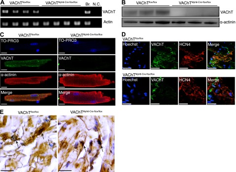
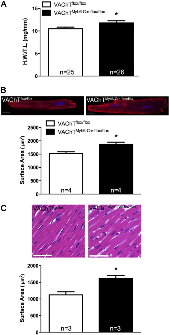
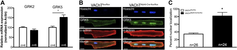
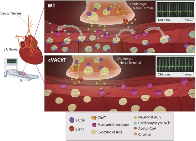
Heart activity and long-term function are regulated by the sympathetic and parasympathetic branches of the nervous system. Parasympathetic neurons have received increased attention recently because acetylcholine (ACh) has been shown to play protective roles in heart disease. However, parasympathetic innervation is sparse in the heart, raising the question of how cholinergic signaling regulates cardiomyocytes. We hypothesized that non-neuronal secretion of ACh from cardiomyocytes plays a role in cholinergic regulation of cardiac activity. To test this possibility, we eliminated secretion of ACh exclusively from cardiomyocytes by targeting the vesicular acetylcholine transporter (VAChT). We find that lack of cardiomyocyte-secreted ACh disturbs the regulation of cardiac activity and causes cardiomyocyte remodeling. Mutant mice present normal hemodynamic parameters under nonstressful conditions; however, following exercise, their heart rate response is increased. Moreover, hearts from mutant mice present increased oxidative stress, altered calcium signaling, remodeling, and hypertrophy. Hence, without cardiomyocyte-derived ACh secretion, hearts from mutant mice show signs of imbalanced autonomic activity consistent with decreased cholinergic drive. These unexpected results suggest that cardiomyocyte-derived ACh is required for maintenance of cardiac homeostasis and regulates critical signaling pathways necessary to maintain normal heart activity. We propose that this non-neuronal source of ACh boosts parasympathetic cholinergic signaling to counterbalance sympathetic activity regulating multiple aspects of heart physiology.

Keywords: VAChT; autonomic function; cardiac hypertrophy; cardiac remodeling; choline acetyltransferase; parasympathetic activity.


Figures

Figure 1.
Figure 1.
Selective elimination of VAChT in cardiomyocytes of cVAChT mice. A–C) VAChT expression was determined by PCR (A; Br., brain, N.C., negative control), immunoblotting (B), and immunofluorescence (C) in adult cardiomyocytes. Scale bars = 25 μm. D) Colabeling for VAChT and HCN4 in whole-mount atrial tissue from control and cVAChT mice. Asterisks indicate VAChT staining in parasympathetic nerve terminals. Scale bars = 25 μm. E) VAChT immunoreactivity in intracardiac parasympathetic ganglia from cVAChT mice. Arrows indicate positive staining for VAChT. Scale bars = 25 μm.
Figure 2.
Figure 2.
A) ACh release from neonatal cardiomyocytes isolated and cultured from control and cVAChT mice. n = number of separate cell isolations for each genotype. ***P < 0.001 vs. control. B) ACh release from control and cVAChT neonatal cardiomyocytes as detected through HPLC with electrochemical detection. n = number of separate cell isolations for each genotype. **P < 0.01 vs. control. C) Bioassay to measure ACh release in cultured neonatal cardiomyocytes using DAF fluorescence. Cells were treated with either carbachol (Carb.) to activate muscarinic receptors and increase NO levels or pyridostigmine (PYR), which preserves secreted ACh, which can then activate muscarinic receptors and increase the production of NO (12). n = number of cells examined from 5 separate cell isolations/genotype. Scale bars = 25 μm. Data are represented as means ± sem. *P < 0.05 vs. control.
Figure 3.
Figure 3.
Analysis of heart rate in cVAChT mice. A) Blood pressure and heart rate analysis in VAChTflox/flox and cVAChT mice using the CODA tail-cuff system. B) Heart rate over 24 h in awake, freely moving VAChTflox/flox and cVAChT mice in their home cage (n≥4 mice/genotype). C) Heart rate response following gentle restraint and i.p. saline injection (n≥4 mice/genotype). D) Heart rate recovery following acute, brief exercise in VAChTflox/flox and cVAChT mice (n≥4 mice/genotype). Data are represented as means ± sem. *P < 0.05 vs. control mice.
Figure 4.
Figure 4.
Cardiac hypertrophy in cVAChT mice. A) Heart weight normalized to tibia length in VAChTflox/flox and cVAChT mice (n=number of mice). B) Surface area of isolated cardiomyocytes (n=number of mice/genotype; ≥75 cells/genotype were analyzed). C) Surface area of cardiomyocytes in situ (n=number of mice/genotype, ≥70 cells/genotype were analyzed). Scale bars = 25 μm. Data are represented as means ± sem. *P < 0.05 vs. control.
Figure 5.
Figure 5.
Cellular stress in cVAChT cardiomyocytes. A) Expression of the cardiac stress markers, β-MHC and ANP, in control and cVAChT cardiomyocytes (n=number of mice). B, C) Immunostaining for ANP in adult cardiomyocytes from control and cVAChT mice. D) Measurement of ROS levels in isolated cardiomyocytes loaded with the MitoSOX superoxide indicator (n=number of cells; ≥3 mice/genotype). E) Measurement of oxidized protein levels in whole hearts from control and cVAChT mice (n=number of mice). F, G) Assessment of calcium transients in isolated ventricular myocytes from VAChTflox/flox and cVAChT mice. F) Representative recordings of line-scan profile of Ca2+ transients in control and cVAChT myocytes. G) Summary of peak Ca2+ (n=number of cardiomyocytes; cells isolated from ≥3 mice/genotype). Scale bars = 25 μm. Data are represented as means ± sem. *P < 0.05 vs. control.
Figure 6.
Figure 6.
Cardiac remodelling in cVAChT cardiomyocytes. A) mRNA expression of GRK5 in isolated cells from control and cVAChT mice (n=number of mice). B, C) Immunostaining for GRK5 in isolated myocytes. Scale bars = 25 μm. Data are represented as means ± sem. *P < 0.05 vs. control.
Figure 7.
Figure 7.
Non-neuronal release of Ach from cardiomyocytes. WT cardiomyocytes secrete ACh in response to increased physiological stress (e.g., exercise) in a VAChT-dependent manner to regulate heart rate. This response is blunted in mice lacking VAChT specifically in cardiomyocytes due to the lack of non-neuronal ACh release.

Comment in

  • Letters to the editor: Cardiomyocyte-secreted acetylcholine.
    Pappano A. Pappano A. FASEB J. 2014 Jan;28(1):1-2. doi: 10.1096/fj.14-0101LTE. FASEB J. 2014. PMID: 24385568 No abstract available.
  • Response.
    Roy A, Fields WC, Rocha-Resende C, Resende RR, Guatimosim S, Prado VF, Gros R, Prado MA. Roy A, et al. FASEB J. 2014 Jan;28(1):2-3. doi: 10.1096/fj.14-0102LTE. FASEB J. 2014. PMID: 24385569 No abstract available.

Similar articles

Cited by

References

    1. Levy M. N. (1997) Neural control of cardiac function. Baillieres Clin. Neurol. 6, 227–244 - PubMed
    1. Kanazawa H., Ieda M., Kimura K., Arai T., Kawaguchi-Manabe H., Matsuhashi T., Endo J., Sano M., Kawakami T., Kimura T., Monkawa T., Hayashi M., Iwanami A., Okano H., Okada Y., Ishibashi-Ueda H., Ogawa S., Fukuda K. (2010) Heart failure causes cholinergic transdifferentiation of cardiac sympathetic nerves via gp130-signaling cytokines in rodents. J. Clin. Invest. 120, 408–421 - PMC - PubMed
    1. Lara A., Damasceno D. D., Pires R., Gros R., Gomes E. R., Gavioli M., Lima R. F., Guimaraes D., Lima P., Bueno C. R., Jr., Vasconcelos A., Roman-Campos D., Menezes C. A., Sirvente R. A., Salemi V. M., Mady C., Caron M. G., Ferreira A. J., Brum P. C., Resende R. R., Cruz J. S., Gomez M. V., Prado V. F., de Almeida A. P., Prado M. A., Guatimosim S. (2010) Dysautonomia due to reduced cholinergic neurotransmission causes cardiac remodeling and heart failure. Mol. Cell. Biol. 30, 1746–1756 - PMC - PubMed
    1. Hoover D. B., Ganote C. E., Ferguson S. M., Blakely R. D., Parsons R. L. (2004) Localization of cholinergic innervation in guinea pig heart by immunohistochemistry for high-affinity choline transporters. Cardiovasc. Res. 62, 112–121 - PubMed
    1. Kawano H., Okada R., Yano K. (2003) Histological study on the distribution of autonomic nerves in the human heart. Heart Vessels 18, 32–39 - PubMed

Publication types

Substances

LinkOut - more resources