. 2013 Oct;123(10):4170-81.
doi: 10.1172/JCI67315. Epub 2013 Sep 9.

Retinal angiogenesis suppression through small molecule activation of p53

Retinal angiogenesis suppression through small molecule activation of p53

Sai H Chavala et al. J Clin Invest. 2013 Oct.

Abstract

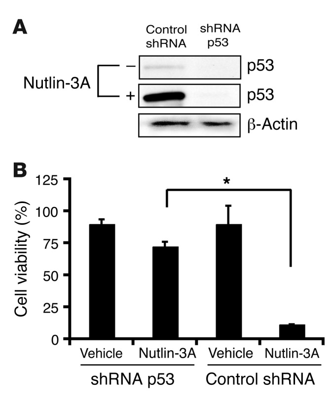
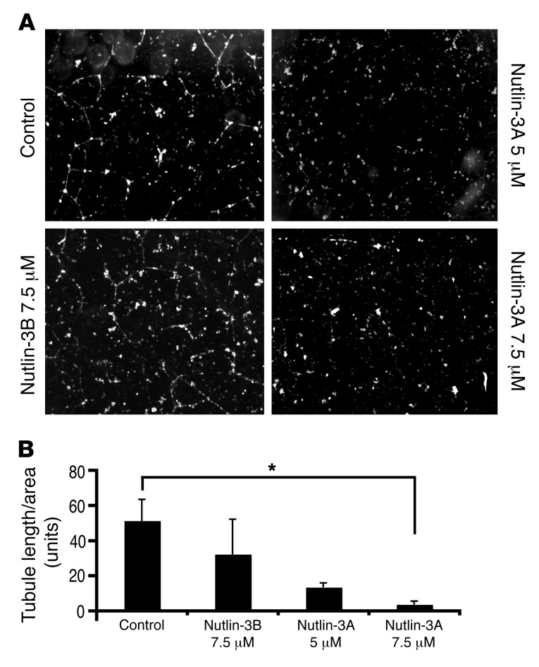
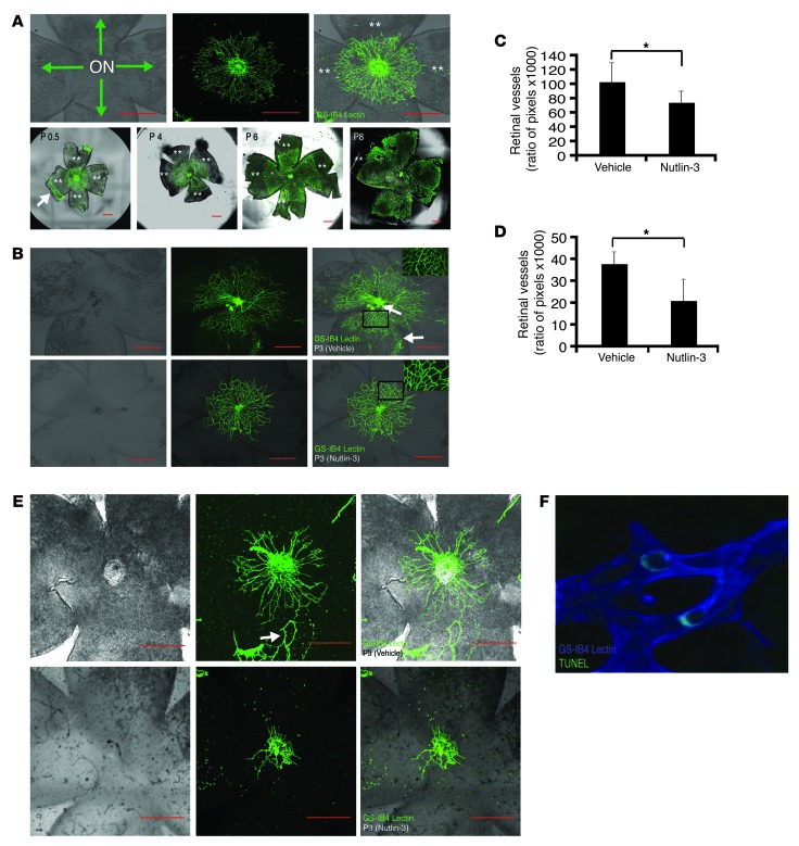
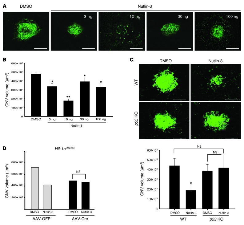
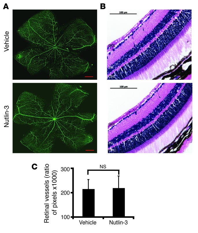
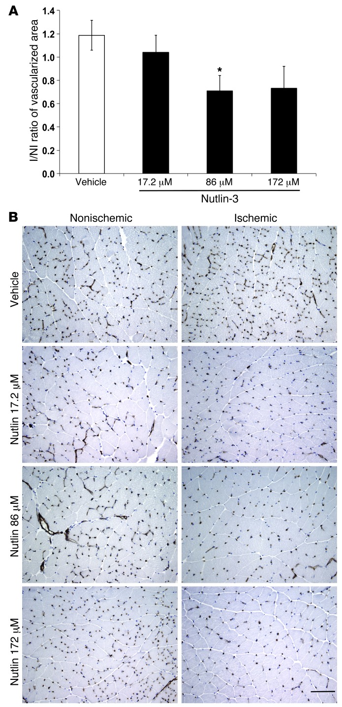
Neovascular age-related macular degeneration is a leading cause of irreversible vision loss in the Western world. Cytokine-targeted therapies (such as anti-vascular endothelial growth factor) are effective in treating pathologic ocular angiogenesis, but have not led to a durable effect and often require indefinite treatment. Here, we show that Nutlin-3, a small molecule antagonist of the E3 ubiquitin protein ligase MDM2, inhibited angiogenesis in several model systems. We found that a functional p53 pathway was essential for Nutlin-3-mediated retinal antiangiogenesis and disruption of the p53 transcriptional network abolished the antiangiogenic activity of Nutlin-3. Nutlin-3 did not inhibit established, mature blood vessels in the adult mouse retina, suggesting that only proliferating retinal vessels are sensitive to Nutlin-3. Furthermore, Nutlin-3 inhibited angiogenesis in nonretinal models such as the hind limb ischemia model. Our work demonstrates that Nutlin-3 functions through an antiproliferative pathway with conceivable advantages over existing cytokine-targeted antiangiogenesis therapies.


Figures

Figure 1
Figure 1. Nutlin-3 inhibits HUVEC proliferation.
(A) Nutlin-3A or Nutlin-3B in 1, 5, or 10 μM concentrations was added to proliferating HUVECs for 36 hours. HUVECs were either unchallenged or challenged with VEGF-A or FGF-2 during the incubation. (B) Cell viability was measured at 12, 24, and 36 hours after incubation. (C) Phase contrast images of representative conditions of serum free, unchallenged HUVECs at 36 hours. Original magnification, ×100.
Figure 2
Figure 2. Nutlin-3 inhibits HUVSMC proliferation.
(A) Confocal immunofluorescence images are displayed characterizing HUVSMCs. The panels indicate that these cells are vimentin (left panel) and smooth muscle actin (middle panel) positive but VE-cadherin (right panel) negative. Original magnification, ×200. (B) Vehicle, Nutlin-3B (Nut-3B) (7.5 μM), or Nutlin-3A (7.5 μM) was added to proliferating HUVSMCs at various time points with FGF-2 or 5% FBS. Various concentrations (0, 7.5, 15, 30 μM) of Nutlin-3A were added to cultures of serum-free, unchallenged HUVSMCs for 24 hours (lower panel). (C) Representative images of serum-free HUVSMCs supplemented with FGF-2 after 72 hours of culture. Original magnification, ×100. Data are mean ± SD and representative of 4 separate experiments performed in at least duplicate. NS, P > 0.05.
Figure 3
Figure 3. Nutlin-3 induces p53 expression and a downstream target in HUVECs and HUVSMCs.
(A) HUVECs were seeded on plastic-bottom culture dishes precoated with gelatin and treated with either 5 μM of Nutlin-3A, 5 μM of Nutlin-3B, or vehicle after 8 hours. Images were taken with an epifluorescent microscope and are representative of p53 expression (middle column) in the nucleus (left column) of HUVECs. Original magnification, ×400. (B) Western blot analysis for p53 and p21 was performed on lysates obtained from HUVECs treated with Nutlin-3A, Nutlin-3B, or vehicle in various concentrations for 8 hours. (C) Representative magnified confocal images are shown of HUVSMCs treated with either 7.5 μM of Nutlin-3A or vehicle after 8 hours. Original magnification, ×200. Nuclear p53 expression is shown in red and TO-PRO-3, a nuclear stain, in blue. (D) Western blot analysis for p53 and p21 was performed on lysates obtained from HUVSMCs treated with Nutlin-3A or vehicle.
Figure 4
Figure 4. Nutlin-3 induces apoptosis in HUVECs but not in HUVSMCs.
(A) Representative HUVEC flow cytometry plots from 3 independent experiments were run in duplicate for 8 hours. (B) TUNEL staining images of HUVECs. Original magnification, ×200. (C) The ratio of the number of TUNEL-positive cells to the number of nuclei found in 5 random fields from each condition are counted by 2 masked observers. Data are mean ± SD and represent 2 independent experiments. (D) qRT-PCR of HUVECs treated for 4 hours. Relative BAX and BCL-2 expression are presented as a ratio. Values are provided as a percentage of DMSO (control) and expressed as mean ± SD. (n = 9 from 3 independent experiments).*P < 0.05. (E) Representative HUVEC flow cytometry plots from 3 independent experiments were run in duplicate for 36 hours. (F) qRT-PCR of BAX and BCL-2 expression in HUVSMCs treated for 4 hours. Values are provided given as a percentage of DMSO (control) ± SD. (n = 12 from 4 independent experiments) NS, P > 0.05. Amount of Nutlin-3A and Nutlin-3B was 7.5 μM and that of Etoposide was 10 μM.
Figure 5
Figure 5. p53 is necessary for Nutlin-3–mediated cell death.
(A and B) HUVECs were infected with a lentiviral vector expressing p53 or control shRNA for 48 hours and then underwent puromycin selection for 72 hours. (A) Cell lysates for Western blot analysis were used to confirm knockdown of p53 in shRNA-infected HUVECs. (B) p53 shRNA– and control shRNA–infected HUVECs were seeded at 1 × 10⁶ cells in 6-well plates and incubated with FGF-2 and either Nutlin-3A (7.5 μM) or vehicle (DMSO) for 48 hours. Cell proliferation was measured at 48 hours by manual counting using trypan blue exclusion. Data represent mean ± SD. *P < 0.05.
Figure 6
Figure 6. Nutlin-3 inhibits capillary tube formation.
(A) HUVECs were seeded on Matrigel matrix and incubated with FGF-2 and 7.5 μM of Nutlin-3A, 5 μM of Nutlin-3A, 7.5 μM of Nutlin-3B, or vehicle (DMSO). The images were taken with an inverted light microscope and are representative of capillary tube formation at 24 hours. Original magnification, ×100. (B) Quantification of Nutlin-3A–mediated capillary tube formation inhibition. Results are expressed as a ratio of tubule length measured to total area examined ± SEM. *P < 0.05.
Figure 7
Figure 7. Nutlin-3 inhibits postnatal retinal vascular proliferation.
(A) Postnatal mouse retinal vascular development after birth (upper left panel). The optic nerve (ON) at the center of a retinal whole mount with green arrows indicating the direction of postnatal blood vessel growth (upper middle and right panels). GS-IB4 lectin staining of a retinal whole mount in the developing mouse pup demonstrating radial growth pattern of postnatal development of retinal vasculature. (lower panels). Double asterisks indicate areas of avascular retina. (B and C) Neonatal mice were given subcutaneous injections in the nape. (B) The retinal vasculature is abrogated in the Nutlin-3–treated eyes (n = 6) (bottom row) compared with the sham-injected mice (n = 4) (middle row). In addition, we noticed loss of smaller caliber vessels (inset, right column) in the Nutlin-3–treated mice. (C) The graph represents retinal vasculature measured as a function of pixels compared with the amount of retinal tissue in each mouse eye (*P < 0.05, representative of 2 independent experiments). (D and E) Neonatal mice were injected in the periocular area of each eye. (E) Nutlin-3–treated (lower row) mice (n = 8) have less retinal vasculature compared with sham-injected mice (n = 8) (upper row). (D) Same quantification method used in B shows a statistically significant difference between the 2 groups (*P < 0.01, representative of 2 independent experiments). (F) Rare TUNEL-positive cells identified in the retinal vasculature of a Nutlin-3–treated mouse. Original magnification, ×400. White arrows point to residual fetal vasculature unable to be removed during dissection. Data represent mean ± SD. *P < 0.05. Scale bars: 500 μm.
Figure 8
Figure 8. Nutlin-3 inhibits pathologic retinal angiogenesis through the p53 pathway.
(A) Stacked confocal image of representative laser-induced CNV lesions in racemic Nutlin-3– and DMSO-treated eyes. Racemic Nutlin-3 showed maximal activity at 10 ng/17.2 μM. (B) Quantification of z-stack confocal images of laser induced CNV in the respective conditions (n = 16–40). (C) Representative images of littermate controls (upper panels) after intravitreal injection of DMSO and racemic Nutlin-3. Similar experiments in p53–/– mice demonstrated no appreciable difference between DMSO- and Nutlin-3–injected mice. (n = 10–13). (D) Hif1aflox/flox mice were given either subretinal AAV1-VMD2-Cre or AAV1-VMD2-GFP. HIF-1α inhibition had no additional impact on Nutlin-3 antiangiogenesis (n = 6–10). Data represent mean ± SEM. *P < 0.05; **P < 0.0001; NS = P > 0.05. P = 0.052 for AAV1-GFP-DMSO vs. AAV1-GFP–Nutlin-3. Scale bars: 100 μm.
Figure 9
Figure 9. Nutlin-3 does not target preexisting blood vessels.
(A and B) Adult mice received a single intravitreal injection of Nutlin-3 or DMSO to investigate the effects of Nutlin-3 on established blood vessels. (A) Confocal images of GS-IB4 lectin–stained and (B) H&E-stained paraffin-embedded sections of retinal vasculature 5 days after injection. Scale bars: 500 μm (A); 100 μm (B).(C) Quantification of retinal vessels shows no difference between sham- (n = 8) and Nutlin-3–injected mice (n = 8) Data represent mean ± SD. NS, P > 0.05, summation of 2 independent experiments.
Figure 10
Figure 10. Nutlin-3 inhibited hind limb ischemia–induced neovascularization.
(A) Quantification of blood capillary density by CD31 immunostaining, expressed as a ratio of ischemic to nonischemic hind limb normalized on myocytes number shows that Nutlin-3 significantly reduced hemangiogenesis when delivered at 86 μM concentration compared with vehicle treatment. At a higher concentration (172 μM), the inhibition was similar but not significant (n = 5). (B) Representative pictures of CD31 immunostaining of capillaries in nonischemic and ischemic muscles of lower limbs revealed a robust neovascular response at 7 days in the vehicle-treated ischemic hind limb. Treatment with Nutlin-3 resulted in significantly reduced CD31 capillary staining in ischemic hind limb muscles. Data represent mean ± SEM. *P < 0.05. Scale bar: 100 μm.

Similar articles

Cited by

References

    1. Rosenfeld PJ, et al. Ranibizumab for neovascular age-related macular degeneration. N Engl J Med. 2006;355(14):1419–1431. doi: 10.1056/NEJMoa054481. - DOI - PubMed
    1. Lux A, Llacer H, Heussen FMA, Joussen AM. Non-responders to bevacizumab (Avastin) therapy of choroidal neovascular lesions. Br J Ophthalmol. 2007;91(10):1318–1322. doi: 10.1136/bjo.2006.113902. - DOI - PMC - PubMed
    1. Dugel PU, et al. Macular epiretinal brachytherapy in treated age-related macular degeneration: MERITAGE study: twelve-month safety and efficacy results. Ophthalmology. 2012;119(7):1425–1431. doi: 10.1016/j.ophtha.2012.01.014. - DOI - PubMed
    1. Petrarca R, Jackson TL. Radiation therapy for neovascular age-related macular degeneration. Clin Ophthalmol. 2011;5:57–63. - PMC - PubMed
    1. Canton VM, et al. 24-Gy low-voltage x-ray irradiation with ranibizumab therapy for neovascular AMD: 6-month safety and functional outcomes. Ophthalmic Surg Lasers Imaging. 2012;43(1):20–24. doi: 10.3928/15428877-20111129-01. - DOI - PubMed

Publication types

MeSH terms