. 2013 Oct;123(10):4423-34.
doi: 10.1172/JCI67867. Epub 2013 Sep 9.

Proximal tubule H-ferritin mediates iron trafficking in acute kidney injury

Proximal tubule H-ferritin mediates iron trafficking in acute kidney injury

Abolfazl Zarjou et al. J Clin Invest. 2013 Oct.

Abstract

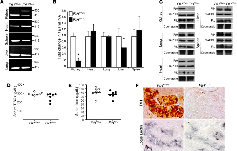
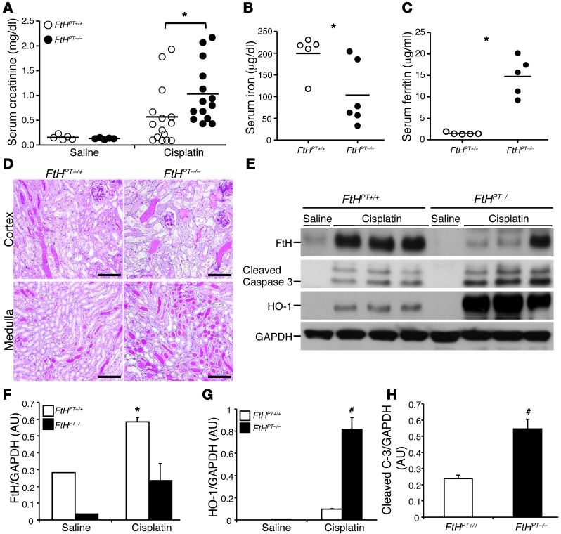
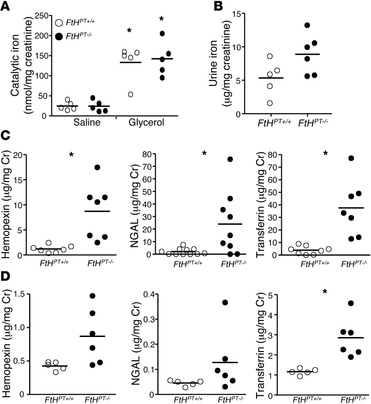
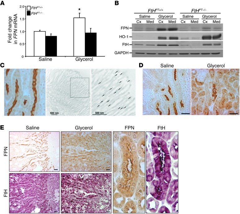
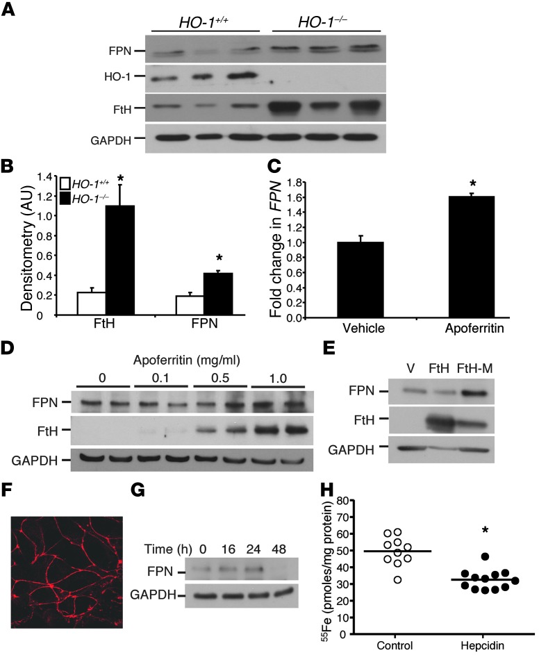
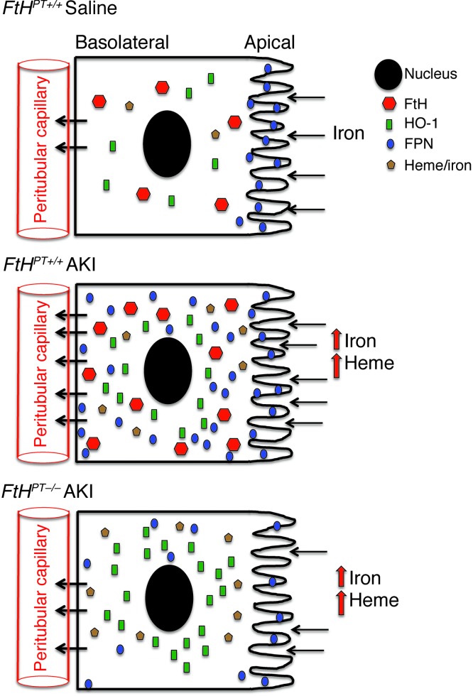
Ferritin plays a central role in iron metabolism and is made of 24 subunits of 2 types: heavy chain and light chain. The ferritin heavy chain (FtH) has ferroxidase activity that is required for iron incorporation and limiting toxicity. The purpose of this study was to investigate the role of FtH in acute kidney injury (AKI) and renal iron handling by using proximal tubule-specific FtH-knockout mice (FtH(PT-/-) mice). FtH(PT-/-) mice had significant mortality, worse structural and functional renal injury, and increased levels of apoptosis in rhabdomyolysis and cisplatin-induced AKI, despite significantly higher expression of heme oxygenase-1, an antioxidant and cytoprotective enzyme. While expression of divalent metal transporter-1 was unaffected, expression of ferroportin (FPN) was significantly lower under both basal and rhabdomyolysis-induced AKI in FtH(PT-/-) mice. Apical localization of FPN was disrupted after AKI to a diffuse cytosolic and basolateral pattern. FtH, regardless of iron content and ferroxidase activity, induced FPN. Interestingly, urinary levels of the iron acceptor proteins neutrophil gelatinase-associated lipocalin, hemopexin, and transferrin were increased in FtH(PT-/-) mice after AKI. These results underscore the protective role of FtH and reveal the critical role of proximal tubule FtH in iron trafficking in AKI.


Figures

Figure 1
Figure 1. Characterization of FtHPT–/– mice.
(A) PCR analysis, performed on genomic DNA from organs, for the presence of the floxed (419 bp) and deleted (530 bp) FtH allele. (B) FtH mRNA expression in the organs of FtHPT+/+ and FtHPT–/– mice, analyzed by real-time PCR. Results were normalized to GAPDH and expressed as fold change compared with FtHPT+/+. Data are mean ± SEM. *P < 0.001 vs. FtHPT+/+. (C) Western blot was performed on organ lysates for FtH expression. Membranes were stripped and reprobed for GAPDH as a loading control. Native gels were run for the detection of FtL, and coomassie stain served as a loading control. (D and E) Total iron-binding capacity (TIBC; D) and total iron levels (E) in the serum of FtHPT+/+ and FtHPT–/– mice. Data are mean ± SEM. No statistical difference was present between groups. (F) Immunohistochemical staining on serial kidney sections from FtHPT+/+ and FtHPT–/– mice for FtH and the proximal tubule marker lotus lectin. For each experiment, n = 4–8 per group.
Figure 2
Figure 2. Ablation of FtH in the proximal tubules worsens heme-mediated AKI.
(A) FtHPT+/+ and FtHPT–/– mice were administered glycerol, and survival was monitored up to 6 days (n = 10 per group). (B) Serum creatinine was measured 24 hours after saline or glycerol administration. Data are mean ± SEM. *P < 0.01 vs. glycerol-treated FtHPT+/+. (C) Total iron levels in the serum of glycerol-treated FtHPT+/+ and FtHPT–/– mice. (D) Serum ferritin was measured 24 hours after saline or glycerol administration. *P < 0.01 vs. glycerol-treated FtHPT+/+. (E) Representative PAS staining of cortex and medulla of glycerol-treated FtHPT+/+ and FtHPT–/– mice. Scale bar: 100 μm. (F and G) FtL and FtH mRNA expression was analyzed by real-time PCR in total RNA extracts of kidneys from saline- or glycerol-treated FtHPT+/+ and FtHPT–/– mice. Data are expressed as fold change in ferritin expression compared with saline-treated FtHPT+/+ mice (n = 3 per group). *P < 0.01. (H) FtH and cleaved caspase-3 expression was verified by Western blot. Blots were stripped and reprobed for GAPDH. (I and J) Expression of the indicated proteins in the kidneys was analyzed by densitometry, normalized to GAPDH, and expressed as mean ± SEM. *P < 0.05 vs. glycerol-treated FtHPT+/+.
Figure 3
Figure 3. Cisplatin nephrotoxicity is exacerbated in the absence of proximal tubule FtH expression.
Mice were administered saline or cisplatin for 3 days (n = 3–10 per group). (A) Serum creatinine, measured at time of harvest and expressed as mg/dl. *P < 0.05 vs cisplatin-treated FtHPT+/+. (B) Total iron levels in the serum of FtHPT+/+ and FtHPT–/– mice after cisplatin administration. *P < 0.05 vs. FtHPT+/+. (C) Serum ferritin in cisplatin-treated FtHPT+/+ and FtHPT–/– mice. *P < 0.01 vs FtHPT+/+. (D) Representative PAS staining of cortex and medulla of saline- or cisplatin-treated FtHPT+/+ and FtHPT–/– mice. Scale bar: 100 μm. (E) FtH, cleaved caspase-3, and HO-1 expression was verified by Western blot. Blots were stripped and reprobed for GAPDH. (FH) Expression of the indicated proteins in the kidneys was analyzed by densitometry, normalized to GAPDH, and expressed as mean ± SEM. *P < 0.05, #P < 0.01 vs. cisplatin-treated FtHPT+/+.
Figure 4
Figure 4. Increased excretion of urinary iron and iron acceptor proteins in FtHPT–/– mice after AKI.
(A) Catalytic iron was measured in the urine of saline- or glycerol-treated FtHPT+/+ and FtHPT–/– mice. Data were normalized to urine creatinine levels and expressed as nanomoles of iron per milligram creatinine. *P < 0.05 vs. respective saline control. (B) Total iron content was measured in the urine of glycerol-treated FtHPT+/+ and FtHPT–/– mice. Data were normalized to urine creatinine and expressed as microgram iron per milligram creatinine. (C and D) Hemopexin, NGAL, and transferrin were measured by ELISA in the urine from glycerol- (C) or cisplatin-treated (D) FtHPT+/+ and FtHPT–/– mice. Data were normalized to urine creatinine and expressed as microgram per milligram creatinine. *P < 0.05 vs. FtHPT+/+.
Figure 5
Figure 5. FtH modulates FPN expression during AKI.
(A) FPN expression in FtHPT+/+ and FtHPT–/– kidneys after saline or glycerol administration, examined by real-time PCR. Data were normalized to GAPDH and expressed as fold change in FPN mRNA relative to saline-treated FtHPT+/+ (n = 3–5 per group). *P < 0.05. (B) FPN, HO-1, and FtH expression in the cortex and medulla of FtHPT+/+ and FtHPT–/– mice, analyzed by Western blot. Membranes were probed for GAPDH as a loading control. (C) Immunohistochemical staining of a kidney section for FPN and immunoelectron micrograph of a proximal tubule in the outer stripe of the outer medulla, illustrating FPN localization to the brush border on the apical surface. Arrows indicate gold particles. (D) Immunohistochemical staining showing differential expression of FPN in the proximal tubules of saline- and glycerol-treated FtHPT+/+ mice. (E) Immunohistochemical staining of serial kidney sections for FPN and FtH after administration of saline or glycerol, as well as magnified views showing their colocalization in the proximal tubule (right, enlarged ×10). Scale bars: 500 nm (C); 500 nm (C, inset); 100 μm (D and E).
Figure 6
Figure 6. FtH regulates FPN expression in renal proximal tubules.
(A) Whole kidney lysates from HO-1+/+ and HO-1–/– mice were analyzed for FPN, HO-1, and FtH levels by Western blot. Membranes were reprobed for GAPDH to demonstrate equal loading. (B) Kidney FPN and FtH expression was analyzed by densitometry analysis, normalized to GAPDH, and expressed as mean ± SEM. *P < 0.05 vs HO-1+/+. (C) FtHPT+/+ proximal tubular epithelial cells were treated in vitro with apoferritin for 8 hours and analyzed for FPN expression by real-time PCR. Data are expressed as fold change relative to vehicle-treated cells. *P < 0.05. (D) Proximal tubular epithelial cells were treated with the indicated doses of apoferritin for 16 hours and analyzed for FPN and FtH expression by Western blot. GAPDH served as a loading control. (E) Proximal tubular epithelial cells were treated with vehicle, FtH, or FtH-M for 16 hours and analyzed for FPN and FtH expression by Western blot. GAPDH served as a loading control. Results are representative of 3 independent experiments. (F) Immunocytochemistry for ZO-1 expression on cells grown on Transwell filters, confirming polarization. Original magnification, ×180. (G) Proximal tubular epithelial cells were treated with hepcidin for the indicated times and analyzed for FPN expression by Western blot. (H) Proximal tubular cells grown on Transwell filters were pretreated with vehicle (control) or hepcidin for 48 hours, 55Fe was administered to the apical compartment for 2 hours, and cellular 55Fe levels were measured and expressed as picomoles of iron per milligram protein. *P < 0.05 vs. control.
Figure 7
Figure 7. Regulation of iron homeostasis by FtH in the proximal tubules of the kidney.
Under basal conditions, FPN is expressed on the apical surface of the proximal tubules, which facilitates iron reabsorption and safe sequestration by ferritin. AKI induces the expression of FtH and HO-1, and FtH in turn increases expression and redistribution of FPN throughout the tubule and toward the basolateral side. In the absence of FtH, FPN induction after AKI is reduced and may potentially impair iron reabsorption. Although HO-1 is substantially increased, these cells are more susceptible to injury and apoptosis, which suggests that HO-1–mediated protection is dependent on FtH.

Comment in

References

    1. Xue JL, et al. Incidence and mortality of acute renal failure in Medicare beneficiaries, 1992 to 2001. J Am Soc Nephrol. 2006;17(4):1135–1142. doi: 10.1681/ASN.2005060668. - DOI - PubMed
    1. Shah SV, Walker PD. Evidence suggesting a role for hydroxyl radical in glycerol-induced acute renal failure. Am J Physiol. 1988;255(3 pt 2):F438–F443. - PubMed
    1. Paller MS. Hemoglobin- and myoglobin-induced acute renal failure in rats: role of iron in nephrotoxicity. Am J Physiol. 1988;255(3 pt 2):F539–F544. - PubMed
    1. Nath KA. Heme oxygenase-1: a provenance for cytoprotective pathways in the kidney and other tissues. Kidney Int. 2006;70(3):432–443. - PubMed
    1. Baliga R, Zhang Z, Baliga M, Ueda N, Shah SV. In vitro and in vivo evidence suggesting a role for iron in cisplatin-induced nephrotoxicity. Kidney Int. 1998;53(2):394–401. doi: 10.1046/j.1523-1755.1998.00767.x. - DOI - PubMed

Publication types

MeSH terms