Review
. 2014 Mar;28(3):473-84.
doi: 10.1038/leu.2013.255. Epub 2013 Sep 10.

Very small embryonic-like stem cells (VSELs) represent a real challenge in stem cell biology: recent pros and cons in the midst of a lively debate

Affiliations
Review

Very small embryonic-like stem cells (VSELs) represent a real challenge in stem cell biology: recent pros and cons in the midst of a lively debate

M Z Ratajczak et al. Leukemia. 2014 Mar.

Abstract

The concept that adult tissue, including bone marrow (BM), contains early-development cells with broader differentiation potential has again been recently challenged. In response, we would like to review the accumulated evidence from several independent laboratories that adult tissues, including BM, harbor a population of very rare stem cells that may cross germ layers in their differentiation potential. Thus, the BM stem cell compartment hierarchy needs to be revisited. These dormant, early-development cells that our group described as very small embryonic-like stem cells (VSELs) most likely overlap with similar populations of stem cells that have been identified in adult tissues by other investigators as the result of various experimental strategies and have been given various names. As reported, murine VSELs have some pluripotent stem cell characteristics. Moreover, they display several epiblast/germline markers that suggest their embryonic origin and developmental deposition in adult BM. Moreover, at the molecular level, changes in expression of parentally imprinted genes (for example, Igf2-H19) and resistance to insulin/insulin-like growth factor signaling (IIS) regulates their quiescent state in adult tissues. In several emergency situations related to organ damage, VSELs can be activated and mobilized into peripheral blood, and in appropriate animal models they contribute to tissue organ/regeneration. Interestingly, their number correlates with lifespan in mice, and they may also be involved in some malignancies. VSELs have been successfully isolated in several laboratories; however, some investigators experience problems with their isolation.


Conflict of interest statement

The authors declare no conflict of interest.

Figures
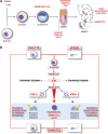
Figure 1
Figure 1
The overall concept of a presence of developmental early epiblast/germline-derived stem cells in adult tissues. (a) Retention of germline potential during ontogenesis. Cells with germline potential are shown in blue. The earliest and the most primitive cell in the germline is the totipotent zygote. The germline potential is subsequently retained during development in Oct-4+ cells located in the inner cell mass cells (ICMs) of the developing blastocyst, epiblast stem cells (EPSCs), primordial germ cells (PGCs) and gonocytes in gonads. Epiblast/germline potential could also be retained in rare early-development small VSELs deposited during development in peripheral tissues as founders for more differentiated monopotent tissue-committed stem cells. Some of these small cells express Oct-4 (shown in blue). (b) The cycle of life—from zygote to germ cells. From a developmental and evolutionary point of view, the germline (shown by red arrows) carries the genome (nuclear and mitochondrial DNA) from one generation to the next, and all somatic cell lines bud out during ontogenesis from the germline to help germline cells accomplish this mission effectively. The germline potential is established in the fertilized oocyte (zygote) and subsequently retained in the morula, inner cell mass of the blastocyst (ICM), EPSC, PGCs and mature germline cells (oocytes and sperm). The first cells that bud out from the germ lineage are trophoectodermal cells, which give rise to the placenta. Subsequently, during gastrulation, EPSCs are a source of PSCs for all three germ layers (meso-, ecto- and endo-derm) and PGCs. We hypothesize that at this stage some EPSCs and PGCs are deposited as Oct-4+ VSELs in tissues developing from meso-, ecto- and endo-derm (blue circles). Blue box and yellow arrows pointing at VSELs indicate mechanism based on epigenetic modification of parentally imprinted genes (for example, at Igf2–H19 and KCNQ1p57Kip2 loci) that keeps these early-development cells quiescent in adult tissues. A similar mechanism based on erasure of imprinting also regulates the quiescent state of PGCs.
Figure 2
Figure 2
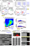
Flow cytometric concerns regarding VSEL identification and isolation. (a) Miyanishi et al. described and subsequently isolated large-size, Syto16-bright, CD45−/int/Lin/Sca-1+ murine cells as ‘VSEL candidates' (lower left plot, blue box), whereas in our opinion, a visible population of smaller, Syto16-dim cells is the most likely VSEL population (lower left plot, red box). Syto16 is not an optimal DNA dye, and its staining also depends on other intracellular components. It is, for instance, visible in the CD45hi/Lin/Sca-1+ population (lower right plot, green box), which is almost a half log unit brighter than the majority of FSChi cells (lower right plot, black box). Moreover, we have a general concern about Sytox-Blue and Sytox16 staining in this study because unfractionated BM cells do not contain any debris or dead cells in such a sample (upper right plot, magenta box), which are visible within subfractions of these cells. (Dot plots were adopted from a paper by Miyanishi et al. (Stem Cell Reports; 2013, 1, 1–11; part of Figure 1C)—published in an open-access journal under Creative Commons Attribution-NonCommercial-No Derivative Works License). (b) Danova-Alt et al., in their recent studies focusing on human VSEL isolation, concluded that CD45/Lin/CD133+ cells (which we consider in fact to be a population enriched in human VSELs) as well as CD45/Lin/CD34+ cells do not exist in CB, whereas such cells are not only found in our samples (lower dot plots, VSELs marked with blue circles), but also in some CB samples (1, 2 and 3) analyzed by Danova-Alt et al. (upper histograms, VSELs marked with blue boxes). These rare stem cell populations were overlooked by Danova-Alt et al. because of their visualization on histograms, but are clearly visible when dot plots are used for analysis. (Histograms were adopted from the paper by Danova-Alt et al. (PLOS One; 2012, 7, e34899; part of Figure 2B)—published in an open-access journal under Creative Commons Attribution License). (c) Szade et al.in their recent studies on VSELs tried to set up a new protocol for murine VSEL isolation. They were unsuccessful, as they focused on an incorrect fraction and, importantly, lost VSELs during an incorrect gating process. A fraction of the very small objects was lost during: (1) gating on the FSC vs SSC plot (left plot, original gate is shown in black, whereas the suggested classical gate for VSELs is shown by a red circle) and (2) excluding doublets (right-hand plots, original gates are shown in black, whereas suggested gates are shown as red boxes, and arrows indicate areas where VSELs were most likely excluded from sorting gates). (Dot plots were adopted from the paper by Szade et al. (PLOS One; 2013, 8, e63329; part of Figure 1A) published in an open-access journal under Creative Commons Attribution License). (d) Images of selected murine cell populations used for size calculation by imaging cytometry. The left panel shows FlowSight-derived brightfield images of CD45−/int/Lin/Sca-1+/Sytox16+ objects that were isolated as ‘VSEL candidates' by Miyanishi et al.. Despite the fact that these cells do not represent the VSEL population described by the Ratajczak group, the quality of images is too poor for accurate quantitative analyses, including size calculations. Importantly, images were collected on the instrument with a low sensitivity that may not be useful for VSEL characterization (scale indicates 20 μm). (Images were adopted from the paper by Miyanishi et al. (Stem Cell Reports; 2013, 1, 1–11; part of Figure S1C)—published in an open-access journal under Creative Commons Attribution-NonCommercial-No Derivative Works License). The middle panel represents size analysis performed by Szade et al. using the ImageStream X system. The histogram shows size analysis of CD45LinSca-1+c-Kit cells, where objects 4-5 μm in size are unfortunately excluded from analysis (as indicated by the orange box), whereas the images are confusingly derived from the distinct CD45LinSca-1+c-Kit+ cell fraction, which does not represent VSELs. Importantly, the quality of images is too poor for accurate data analysis. (Data were adopted from the paper by Szade et al. (PLOS One; 2013, 8, e63329; part of Figure S2)—published in an open-access journal under Creative Commons Attribution License). The right panel shows representative images of murine VSELs (CD45/Lin/Sca-1+/7-AAD+ cells), HSCs (CD45+/Lin/Sca-1+/7-AAD+ cells) and platelets (CD41+/7-AAD objects) obtained with the ImageStream 100 system and used for VSEL size analysis. Importantly, with an optimized instrument and good-quality images, even objects as small as 2-μm platelets may be visible and quantified by IDEAS software. Cyan areas show masking used for calculations of cell size by Zuba-Surma et al.
Figure 3
Figure 3
Classical sorting strategy for murine BM-derived VSEL isolation by fluorescence-activated cell sorting (FACS). Agranular, small events ranging from 2 to 10 μm (as initially set up with Flow Cytometry Size beads, Invitrogen/Molecular Probes) are included in an ‘extended lymphgate' on an FSC vs SSC dot plot (region R1; a). The population of cells from region R1 may be additionally depleted of doublets (gate R2; b) to enhance sorting purity (b). The single-cell fraction from gate R2 is further analyzed for Sca-1 and Lin expression and exclusively Lin/Sca-1+ cells are gated (R3) to avoid erythroblast contamination (c). The population from region R3 is subsequently separated into CD45 and CD45+ subpopulations visualized in regions R4 and R5, respectively, on a CD45 vs SSC dot plot (d). CD45-dim objects are preferentially included in the CD45+ population (region R5). If BM cells are gated strictly according to these steps (with special caution for R1, R3, R4 and R5 gate set-up), the populations of VSELs and HSCs derived from regions R4 and R5, respectively, separate cleanly when ‘back-gated' on FSC vs SSC dot plots (e). Both stem cell fractions may be additionally purified by final gating, including size-related regions R6 and R7 for VSEL and HSC sorting, respectively. Percentages represent the content of each fraction in the representative murine BM sample.

Similar articles

Cited by

References

    1. Tachibana M, Amato P, Sparman M, Gutierrez NM, Tippner-Hedges R, Ma H et al. Human embryonic stem cells derived by somatic cell nuclear transfer. Cell 2013; 153: 1228–1238. - PMC - PubMed
    1. Takahashi K, Yamanaka S. Induction of pluripotent stem cells from mouse embryonic and adult fibroblast cultures by defined factors. Cell 2006; 126: 663–676. - PubMed
    1. Zhao T, Zhang ZN, Rong Z, Xu Y. Immunogenicity of induced pluripotent stem cells. Nature 2011; 474: 212–215. - PubMed
    1. Park IH, Zhao R, West JA, Yabuuchi A, Huo H, Ince TA et al. Reprogramming of human somatic cells to pluripotency with defined factors. Nature 2008; 451: 141–146. - PubMed
    1. Lee AS, Tang C, Rao MS, Weissman IL, Wu JC. Tumorigenicity as a clinical hurdle for pluripotent stem cell therapies. Nat Med 2013; 19: 998–1004. - PMC - PubMed

Publication types