Review
. 2013 Nov 1;591(21):5279-90.
doi: 10.1113/jphysiol.2013.256495. Epub 2013 Sep 9.

Calcium signalling of human pluripotent stem cell-derived cardiomyocytes

Affiliations
Review

Calcium signalling of human pluripotent stem cell-derived cardiomyocytes

Sen Li et al. J Physiol. .

Abstract

Loss of cardiomyocytes (CMs), which lack the innate ability to regenerate, due to ageing or pathophysiological conditions (e.g. myocardial infarction or MI) is generally considered irreversible, and can lead to conditions from cardiac arrhythmias to heart failure. Human (h) pluripotent stem cells (PSCs), including embryonic stem cells (ESC) and induced pluripotent stem cells (iPSCs), can self-renew while maintaining their pluripotency to differentiate into all cell types, including CMs. Therefore, hPSCs provide a potential unlimited ex vivo source of human CMs for disease modelling, drug discovery, cardiotoxicity screening and cell-based heart therapies. As a fundamental property of working CMs, Ca(2+) signalling and its role in excitation-contraction coupling are well described. However, the biology of these processes in hPSC-CMs is just becoming understood. Here we review what is known about the immature Ca(2+)-handling properties of hPSC-CMs, at the levels of global transients and sparks, and the underlying molecular basis in relation to the development of various in vitro approaches to drive their maturation.


Figures

Figure 1
Figure 1. Schematic comparison of Ca2+ signalling pathways in adult and hPSC-derived cardiomyocytes
In both adult (A) and hPSC-CMs (B), Ca2+ entry via ICaL triggers Ca2+ release from SR via RyRs, leading to the rise of Ca2+ transient; the subsequent decay is similarly accomplished by Ca2+ reuptake and extrusion via SERCA and NCX, respectively. The smaller amplitude and slower kinetics and null inotropic response of hPSC-CMs compared to adult can be attributed to the following differences: (1) lack of junction (JCTN) and triadin (TRDN) to facilitate RyR function in hPSC-CMs; (2) lack of calsequestrin (CSQ) for SR Ca buffering (rather, calreticulin (CALR) is expressed in hPSC-CMs); (3) lack of phospholamban (PLB) for sarcoplasmic–endoplasmic reticulum Ca2+-ATPase (SERCA) modulation; (4) lower SERCA and RyR expression in hPSC-CMs; (5) lack of T-tubules which contributes to a U-shape of Ca2+ propagation wavefront.
Figure 2
Figure 2
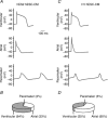
Action potentials of ventricular, atrial and pacemaker cardiomyocytes derived from HES2 (A) and H1 hESCs (C). The corresponding pie charts (B and D). Adapted from Moore et al. (2008).
Figure 3
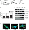
Figure 3. Functional yet immature Ca2+ handling in hESC-CMs compared with fetal ventricular CMs (FLV-CMs) and adult ventricular CMs (ALV-CMs)
A, caffeine-induced Ca2+ transient in HES2-CMs, H1-CMs and FLV-CMs. B, percentage of respective CMs that are sensitive to caffeine treatment. C, Ca2+-handling protein expression profile in HES2-CMs, H1-CMs, FLV-CMs and ALV-CMs. D, immunostaining of ryanodine receptors in HES2-CMs, H1-CMs and FLV-CMs. Adapted from Liu et al. (2007).
Figure 4
Figure 4. Absence of T-tubules in hESC-CMs
Di-8-ANEPPS staining shows no intracellular fluorescent spots in hESC-CMs (A) compared with adult cardiomyocytes (C). B and D, Atomic force microscopy imaging reveals no T-tubules, present as regularly spaced pores, in hESC-CMs. E, lack of T-tubules results in a non-uniform, U-shaped Ca2+ transient. Adapted from Lieu et al. (2009).
Figure 5
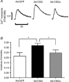
Figure 5. CSQ overexpression increases the amplitude of caffeine-induced Ca2+ transient
A, representative caffeine-induced Ca2+ transient tracings for Ad-GFP, Ad-CSQ and Ad-CSQΔ transduced hESC-CMs. B, bar graphs of amplitude. *P < 0.05. Adapted from Liu et al. (2009).
Figure 6
Figure 6. Adenovirus-mediated Kir2.1 overexpression led to hESC-CMs maturation
A, representative tracings of human embryonic stem cell-derived cardiomyocytes produced by directed differentiation protocol (ddhESC-CMs), showing action potentials (APs) of spontaneously firing (left) and quiescent (right) ventricular CMs. Arrow indicates phase 4-like depolarization. B, APs of Ad-Kir2.1-transduced ventricular ddhESC-CMs. Ik1 in inset. The phase 4-like depolarization was eliminated (arrow) by Ad-Kir2.1 transduction (left). The percentage of quiescent ventricular ddhESC-CMs increased significantly to 100% after Ad-Kir2.1 transduction (middle). Resting membrane potentials (RMPs) of Ad-Kir2.1-transduced ventricular ddhESC-CMs became significantly hyperpolarized relative to control (right). C, after Ad-Kir2.1 transduction, the mRNA expression of contractile elements is significantly reduced relative to control hESC-CMs. D, both electrically conditioned atrial and ventricular ddhESC-CMs harboured action potentials without phase 4-depolarization (left). More hyperpolarized resting membrane potential (RMP) was shown (right). E, caffeine-elicited Ca2+ transients from control (blue) and electrically conditioned (red) ddhESC-CMs, and their averaged peak amplitude were also presented in bar graph. Adapted from Lieu et al. (2012).

Similar articles

Cited by

References

    1. Anderson D, Self T, Mellor IR, Goh G, Hill SJ, Denning C. Transgenic enrichment of cardiomyocytes from human embryonic stem cells. Mol Ther. 2007;15:2027–2036. - PubMed
    1. Awasthi S, Matthews DL, Li RA, Chiamvimonvat N, Lieu DK, Chan JW. Label-free identification and characterization of human pluripotent stem cell-derived cardiomyocytes using second harmonic generation (SHG) J Microscopy Biophoton. 2012;5:57–66. - PMC - PubMed
    1. Bassani JWM, Yuan WL, Bers DM. Fractional SR Ca release is regulated by trigger Ca and SR Ca content in cardiac myocytes. Am J Physiol Cell Physiol. 1995;268:C1313–C1319. - PubMed
    1. Beard NA, Laver DR, Dulhunty AF. Calsequestrin and the calcium release channel of skeletal and cardiac muscle. Prog Biophys Mol Biol. 2004;85:33–69. - PubMed
    1. Bers DM. Cardiac excitation–contraction coupling. Nature. 2002;415:198–205. - PubMed

Publication types

MeSH terms

LinkOut - more resources