. 2014 Nov;53(11):907-16.
doi: 10.1002/mc.22050. Epub 2013 Sep 9.

Role of mitogen-activated protein kinases and Mcl-1 in apoptosis induction by withaferin A in human breast cancer cells

Affiliations

Role of mitogen-activated protein kinases and Mcl-1 in apoptosis induction by withaferin A in human breast cancer cells

Eun-Ryeong Hahm et al. Mol Carcinog. 2014 Nov.

Abstract

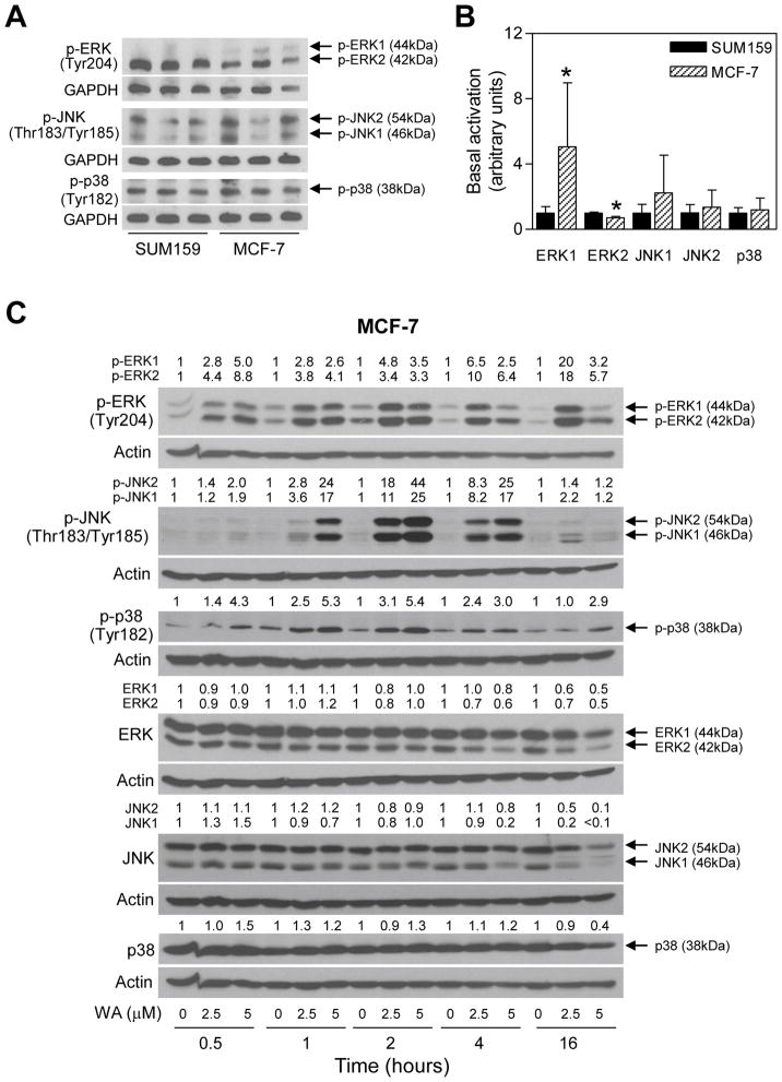
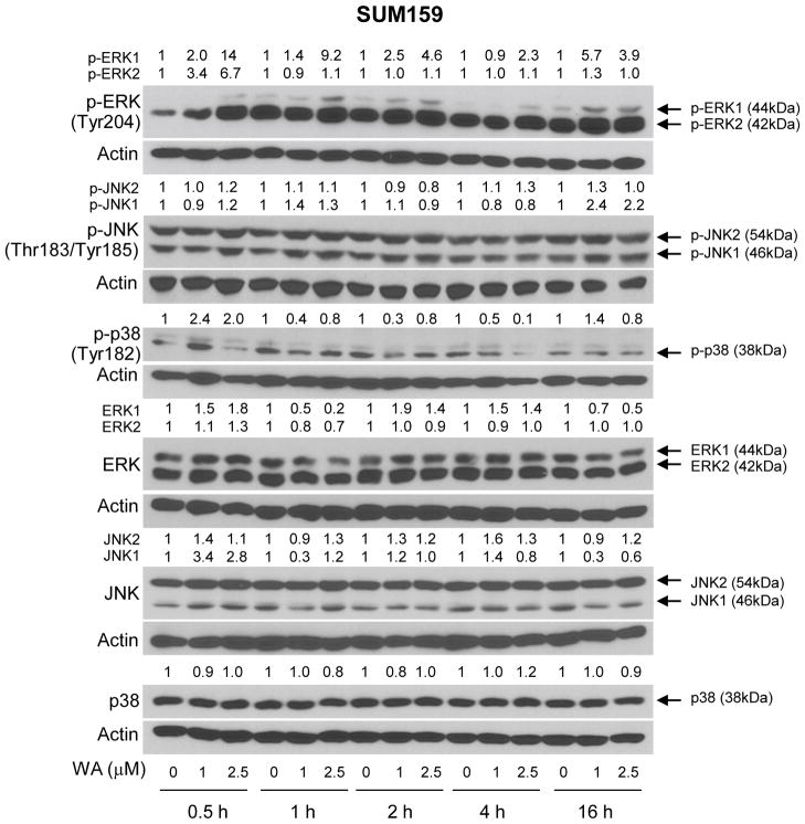
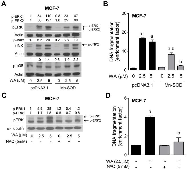
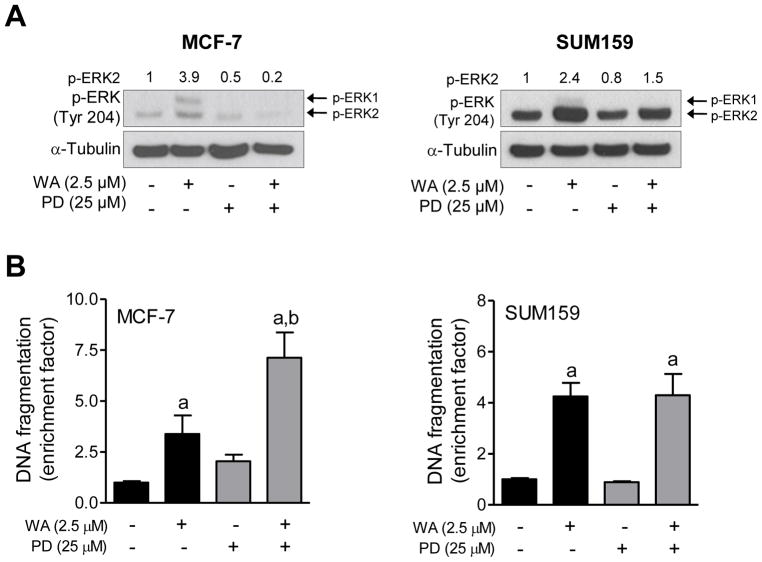
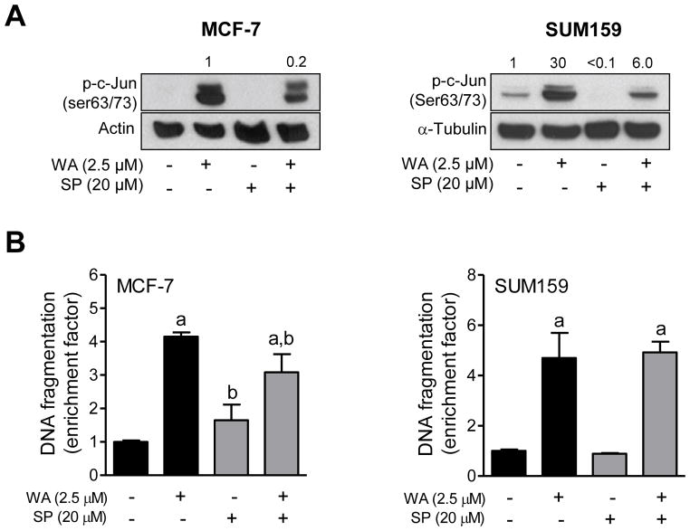
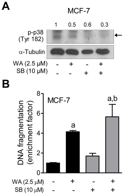
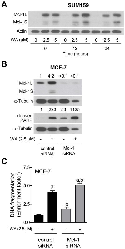
Withaferin A (WA), a bioactive constituent of Ayurvedic medicine plant Withania somnifera, is a potent apoptosis inducer in cancer cells but the mechanism of cell death induction is not fully characterized. The present study was undertaken to determine the role of mitogen-activated protein kinases (MAPK), including c-jun NH2 -terminal kinase (JNK), extracellular signal-regulated kinase (ERK), and p38 MAPK, and anti-apoptotic protein myeloid cell leukemia-1 (Mcl-1) in regulation of WA-induced apoptosis using human breast cancer cells. Exposure of MCF-7 (estrogen responsive) and SUM159 (triple negative) human breast cancer cells to WA resulted in increased phosphorylation of ERK, JNK, and p38 MAPK, but these effects were relatively more pronounced in the former cell line than in SUM159. Overexpression of manganese-superoxide dismutase conferred partial protection against WA-mediated hyperphosphorylation of ERK, but not JNK or p38 MAPK. Cell death resulting from WA treatment in MCF-7 cells was significantly augmented by pharmacological inhibition of ERK and p38 MAPK. Interestingly, the WA-induced apoptosis in MCF-7 cells was partially but significantly blocked in the presence of a JNK-specific inhibitor. Pharmacological inhibition of ERK or JNK had no effect on WA-induced apoptosis in SUM159 cells. The WA-treated cells exhibited induction of long and short forms of Mcl-1. RNA interference of Mcl-1 alone triggered apoptosis. Furthermore, the WA-induced cell death in MCF-7 cells was modestly but significantly augmented by knockdown of the Mcl-1 protein. These observations indicate that: MAPK have cell line-specific role in cell death by WA, and Mcl-1 induction confers modest protection against WA-induced apoptosis.

Keywords: MAPK; Mcl-1; apoptosis; breast cancer; withaferin A.


Figures

Figure 1
Figure 1
WA treatment activates MAPK in MCF-7 human breast cancer cells. (A–B) Determination of basal activation of MAPK in MCF-7 and SUM159 cells. The cells (5×10⁵ cells per dish) were plated in triplicate in 6-cm dish. After overnight incubation, the cells were collected and subjected to western blotting for phosphorylated forms of ERK, JNK, and p38 MAPK. Quantitation of basal level of phosphorylated MAPK in MCF-7 and SUM159 from two-independent experiments is shown in the bar graph. Results shown are mean S.D. (n = 6). Statistical significance (*, P<0.05) of difference in expression level between MCF-7 and SUM159 cells was analyzed by unpaired Student’s t-test. (C) Effect of WA treatment on activation of MAPK in MCF-7 cells. Lysates from MCF-7 cells treated with the indicated concentrations of WA for specified time periods were subjected to western blotting for phosphorylated and total forms of ERK, JNK, and p38. The number on top of the band represents fold change in level compared to corresponding DMSO-treated control.
Figure 2
Figure 2
WA treatment activates MAPK in SUM159 cells. Effect of WA treatment on activation of MAPK in SUM159 cells. Lysates from SUM159 cells treated with the indicated concentrations of WA for specified time periods were subjected to western blotting for phosphorylated and total forms of ERK, JNK, and p38 MAPK. The number on top of the band represents fold change in level compared to corresponding DMSO-treated control.
Figure 3
Figure 3
ROS are involved in apoptosis by WA in MCF-7 cells. (A) Effect of overexpression of Mn-SOD on WA-mediated activation of MAPK. The MCF-7 cells stably transfected with pcDNA3.1 empty vector or pcDNA3.1 vector encoding for Mn-SOD were treated with the desired concentrations of WA for 2 hours. The total lysates from these cells were subjected to western blotting for phosphorylated MAPK. Quantitation relative to DMSO-treated empty vector transfected cells is shown. (B) Detection of apoptosis (histone-associated DNA fragment release into the cytosol) in MCF-7 cells stably transfected with pcDNA3.1 empty vector or pcDNA3.1 vector encoding for Mn-SOD. The cells were treated with the indicated doses of WA for 24 h and then processed for apoptosis assay. Statistical significance (P<0.05) was analyzed one-way ANOVA with Bonferroni’s multiple comparison test. aSignificantly different (P<0.05) compared with each control. bSignificantly different (P<0.05) between groups with the same dose. The results shown are mean ± S.D (n= 3). (C) Effect of NAC on WA-mediated activation of ERK. MCF-7 cells (5×10⁵ cells per dish) were plated in 6-cm dish and allowed to attach. After overnight incubation, the cells were pretreated with 5 mM NAC for 2 h and then treated with indicated doses of WA in the absence or presence of NAC for 2 h. After treatment, the cells were collected and processed for immunoblotting. (D) Effect of NAC on WA-mediated apoptosis induction in MCF-7 cells. The cells were pretreated with 5 mM NAC for 2 h and then treated with 2.5 μM WA in the absence or presence of NAC for 24 h. Statistical significance (P<0.05) was analyzed by one-way ANOVA with Bonferroni’s multiple comparison test. aSignificantly different (P<0.05) compared with each control. bSignificantly different (P<0.05) between groups with the same dose. The results shown are mean ± S.D. (n= 2–3).
Figure 4
Figure 4
The effect of pharmacological inhibition of ERK on WA-mediated apoptosis in human breast cancer cells. The MCF-7 and SUM159 human breast cancer cells were pretreated with 25 μM PD98059 for 1 h, and then exposed to 2.5 μM WA in the absence or presence of PD98059 for an additional 24 h. (A) Western blotting for phosphorylated ERK. Quantitation relative to DMSO-treated cells is shown. (B) Histone-associated DNA fragment release into the cytosol. Quantitation relative to DMSO-treated cells is shown. Combined results (n = 6) from two independent experiments are shown as mean ± S.D. Statistical significance was determined by one-way ANOVA with Bonferroni’s multiple comparison test. aSignificantly different (P<0.05) compared with respective control. bSignificantly different (P<0.05) between groups at the same dose.
Figure 5
Figure 5
The effect of pharmacological inhibition of JNK on WA-mediated apoptosis in human breast cancer cells. The MCF-7 and SUM159 human breast cancer cells were pretreated with 20 μM SP600125 for 1 h, exposed to 2.5 μM WA in the absence or presence of SP600125 for 24 h, and then processed for western blot analysis or apoptosis detection. (A) Western blotting for phosphorylated c-jun. Quantitation relative to DMSO-treated cells is shown. (B) Histone-associated DNA fragment release into the cytosol. Quantitation relative to DMSO-treated cells is shown. Combined results (n = 6) from two independent experiments are shown as mean ± S.D. Statistical significance was determined by one-way ANOVA with Bonferroni’s multiple comparison test. aSignificantly different (P<0.05) compared with respective control. bSignificantly different (P<0.05) between groups at the same dose.
Figure 6
Figure 6
The effect of pharmacological inhibition of p38 MAPK on WA-mediated apoptosis in MCF-7 cells. The MCF-7 cells were pretreated with 10 μM SB202190 for 1 h, exposed to 2.5 μM WA in the absence or presence of SB202190 for 24 h, and then processed for western blot analysis or apoptosis detection. (A) Western blotting for phosphorylated p38 MAPK and cleaved PARP. Quantitation for phosphorylated p38MAPK relative to DMSO-treated cells is shown. (B) Histone-associated DNA fragment release into the cytosol. Quantitation relative to DMSO-treated cells is shown. Combined results (n = 6) from two independent experiments are shown as mean ± S.D. Statistical significance was determined by one-way ANOVA with Bonferroni’s multiple comparison test. aSignificantly different (P<0.05) compared with respective control. bSignificantly different (P<0.05) between groups at the same dose.
Figure 7
Figure 7
The effect of Mcl-1 knockdown on WA-mediated apoptosis in MCF-7 cells. (A) Western blot analysis for Mcl-1 using lysates from SUM159 cells treated with the indicated doses of WA for specified time periods. The number on top of the immunoreactive band represents change of protein expression level relative to corresponding DMSO-treated control. (B) Western blotting for Mcl-1 and cleaved PARP using lysates from transiently transfected MCF-7 cells with a non-specific control siRNA or a Mcl-1-targeted siRNA and treated with 2.5 μM WA for 24 h. Quantitation relative to DMSO-treated control siRNA transfected cells is shown. (C) Histone-associated DNA fragment release into the cytosol in MCF-7 cells transiently transfected with a non-specific control siRNA or a Mcl-1 targeted siRNA and treated with 2.5 μM WA for 24 h. Quantitation relative to DMSO-treated control siRNA transfected cells is shown. Combined results (n = 6) from two independent experiments are shown as mean ± S.D. Statistical significance was determined by one-way ANOVA with Bonferroni’s multiple comparison test. aSignificantly different (P<0.05) compared with respective control. bSignificantly different (P<0.05) between groups at the same dose.

References

    1. Garodia P, Ichikawa H, Malani N, Sethi G, Aggarwal BB. From ancient medicine to modern medicine: Ayurvedic concepts of health and their role in inflammation and cancer. J Soc Integr Oncol. 2007;5:25–37. - PubMed
    1. Gupta SK, Mohanty I, Talwar KK, et al. Cardioprotection from ischemia and reperfusion injury by Withania somnifera: A hemodynamic, biochemical and histopathological assessment. Mol Cell Biochem. 2004;260:39–47. - PubMed
    1. Ahmad M, Saleem S, Ahmad AS, et al. Neuroprotective effects of Withania somnifera on 6-hydroxydopamine induced Parkinsonism in rats. Hum Exp Toxicol. 2005;24:137–147. - PubMed
    1. Devi PU, Sharada AC, Solomon FE. Antitumor and radiosensitizing effects of Withania somnifera (Ashwagandha) on a transplantable mouse tumor, Sarcoma-180. Indian J Exp Biol. 1993;31:607–611. - PubMed
    1. Widodo N, Kaur K, Shrestha BG, et al. Selective killing of cancer cells by leaf extract of Ashwagandha: Identification of a tumor-inhibitory factor and the first molecular insights to its effect. Clin Cancer Res. 2007;13:2298–2306. - PubMed

Publication types

MeSH terms