. 2013 Dec;41(22):10150-6.
doi: 10.1093/nar/gkt773. Epub 2013 Sep 9.

Transcriptional control by two interacting regulatory proteins: identification of the PtxS binding site at PtxR

Affiliations

Transcriptional control by two interacting regulatory proteins: identification of the PtxS binding site at PtxR

Abdelali Daddaoua et al. Nucleic Acids Res. 2013 Dec.

Abstract

The PtxS and PtxR regulators control the expression of the glucose dehydrogenase genes from the Pgad promoter in Pseudomonas aeruginosa. These regulators bind to their cognate operators, that are separated by ∼50 nt, within the promoter region and interact with each other creating a DNA-loop that prevents RNA polymerase promoter access. Binding of the 2-ketogluconate effector to PtxS caused PtxS/PtxR complex dissociation and led to the dissolution of the repression loop facilitating the entry of the RNA polymerase and enabling the transcription of the gad gene. We have identified a hydrophobic surface patch on the PtxR putative surface that was hypothesized to correspond to the binding site for PtxS. Two surface-exposed residues in this patch, V173 and W269, were replaced by alanine. Isothermal titration calorimetry assays showed that PtxS does not interact with the mutant variants of PtxR. Electrophoretic mobility shift assay and DNAase I footprinting assays proved that both regulators bind to their target operators and that failure to interact with each other prevented the formation of the DNA-loop. In vitro transcription showed that PtxS per se is sufficient to inhibit transcription from the Pgad promoter, but that affinity of PtxS for its effector is modulated by PtxR.


Figures

Figure 1.
Figure 1.
Homology model of PtxR. This model was generated using the CrgA structure of Neisseria meningitidis [pdb: 3hhg, and (14)] as template. Perpendicular view to potential dimerization interface. (A) Surface potential of the model. Highlighted is a hydrophobic patch. Red colour indicates negative charges, blue positive charges and grey uncharged residues. (B) Ribbon diagram of the model at the same orientation. Amino acids W269 and V173, which were replaced by alanine residues, are highlighted.
Figure 2.
Figure 2.
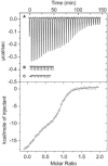
Microcalorimetric binding studies of PtxS to native and mutant PtxR. In all experiments, 3 µM of native or mutant PtxR was titrated with 3.2 µl aliquots of 50 mM of PtxS. (A) Titration of wild-type PtxR and PtxS. (B) Titration of PtxR(V173A) with PtxS. (C) Titration of PtxR(W629A) with PtxS. Top: Titration raw data. Bottom: Integrated and dilution corrected raw data for the titration of PtxR with PtxS. Data were fitted with the ‘Two binding site model’ of the MicroCal version of ORIGIN.
Figure 3.
Figure 3.
Evaluation of the change in electrophoretic mobility of Pgad DNA following binding to PtxS/PtxR or PtxS/mutant PtxR. EMSAs were conducted using 20 µM of purified protein with 2 nM of the Pgad DNA end-labelled with [γ-32P] [from left to right: Free DNA, PtxS(WT), PtxR(WT), PtxR(V173A), PtxR(W269A), PtxS(WT)/PtxR(WT), PtxS(WT)/PtxR(V173A) and PtxS(WT)/PtxR(W269A)].
Figure 4.
Figure 4.
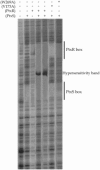
DNase I footprinting assays of promoter Pgad. Experiments were conducted as described in ‘Materials and Methods’ section. (A) Lane 1: free DNA, lane 2: DNA + 10 µM PtxS, lane 3: DNA + 10 µM PtxR, lane 4: DNA + 10 µM PtxS and PtxR, lane 5: DNA + 20 mM PtxS and PtxR, lane 6: DNA + 20 µM PtxS and 20 µM PtxR(V173A) and lane 7: DNA + 20 µM PtxS and 20 µM PtxR(W269A).
Figure 5.
Figure 5.
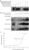
In vitro transcription from Pgad. Transcription assays were carried out as described in ‘Materials and Methods’ section. (A) The assay performed in the presence of 20 μM combined wild-type PtxS with wild type PtxR, PtxR(V173A) or PtxR(W269A) mutants. (B) In vitro transcription assays obtained by supplementation of increase concentration of 2-ketogluconate (0–150 µM). (C) The densitometric analysis of the in vitro transcription gels. Circles: WT PtxR protein, triangle: V173A PtxR protein and star: W269A.

Similar articles

Cited by

References

    1. Cao H, Baldini RL, Rahme LG. Common mechanisms for pathogens of plants and animals. Annu. Rev. Phytopathol. 2001;39:259–284. - PubMed
    1. Rahme LG, Stevens EJ, Wolfort SF, Shao J, Tompkins RG, Ausubel FM. Common virulence factors for bacterial pathogenicity in plants and animals. Science. 1995;268:1899–1902. - PubMed
    1. Iglewski BH, Kabat D. NAD-dependent inhibition of protein synthesis by Pseudomonas aeruginosa toxin. Proc. Natl Acad. Sci. USA. 1975;72:2284–2288. - PMC - PubMed
    1. Hamood AN, Colmer HJ, Carty NL. Regulation of Pseudomonas aeruginosa exotoxin A synthesis. In: Ramos JL, editor. Pseudomonas: Virulence and Gene Regulation. Vol. 2. Amsterdam: Plenum Press; 2004. pp. 389–423.
    1. Vasil ML, Ochsner UA, Johnson Z, Colmer JA, Hamood AN. The fur-regulated gene encoding the alternative sigma factor PvdS is required for iron-dependent expression of the LysR-type regulator ptxR in Pseudomonas aeruginosa. J. Bacteriol. 1998;180:6784–6788. - PMC - PubMed

Publication types

MeSH terms