. 2013 Sep 24;110(39):15650-5.
doi: 10.1073/pnas.1315006110. Epub 2013 Sep 9.

CYP2R1 is a major, but not exclusive, contributor to 25-hydroxyvitamin D production in vivo

Affiliations

CYP2R1 is a major, but not exclusive, contributor to 25-hydroxyvitamin D production in vivo

Jinge G Zhu et al. Proc Natl Acad Sci U S A. .

Abstract

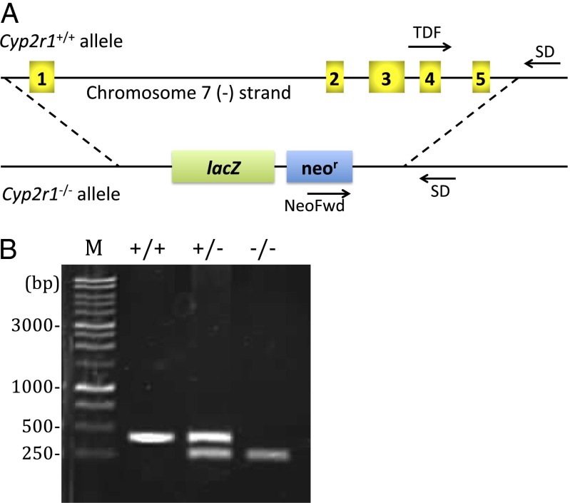
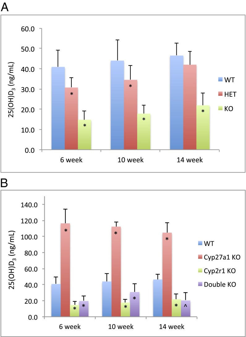
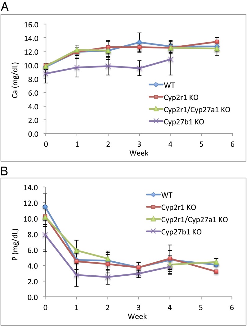
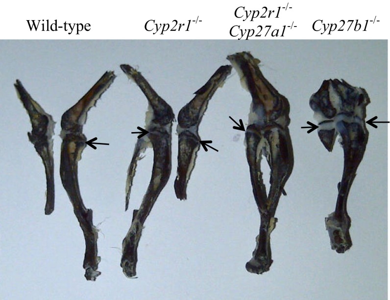
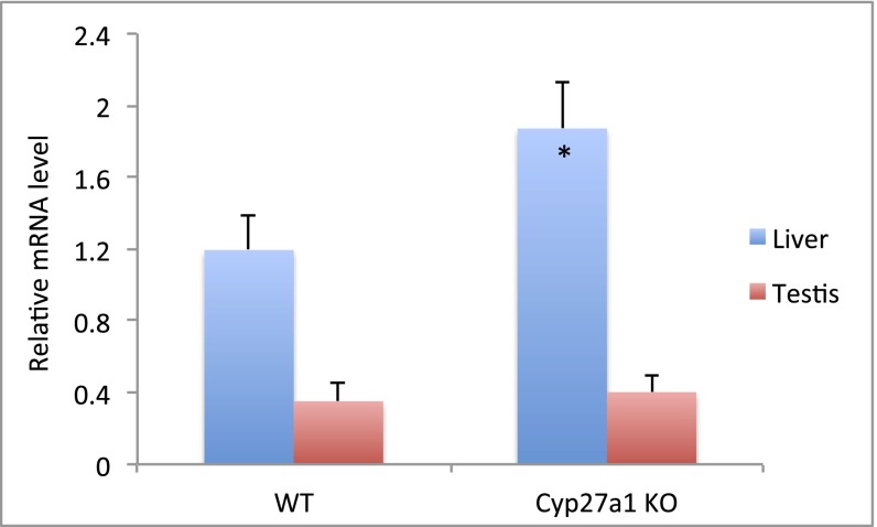
Bioactivation of vitamin D consists of two sequential hydroxylation steps to produce 1α,25-dihydroxyvitamin D3. It is clear that the second or 1α-hydroxylation step is carried out by a single enzyme, 25-hydroxyvitamin D 1α-hydroxylase CYP27B1. However, it is not certain what enzyme or enzymes are responsible for the initial 25-hydroxylation. An excellent case has been made for vitamin D 25-hydroxylase CYP2R1, but this hypothesis has not yet been tested. We have now produced Cyp2r1 (-/-) mice. These mice had greater than 50% reduction in serum 25-hydroxyvitamin D3. Curiously, the 1α,25-dihydroxyvitamin D3 level in the serum remained unchanged. These mice presented no health issues. A double knockout of Cyp2r1 and Cyp27a1 maintained a similar circulating level of 25-hydroxyvitamin D3 and 1α,25-dihydroxyvitamin D3. Our results support the idea that the CYP2R1 is the major enzyme responsible for 25-hydroxylation of vitamin D, but clearly a second, as-yet unknown, enzyme is another contributor to this important step in vitamin D activation.


Conflict of interest statement

The authors declare no conflict of interest.

Figures

Fig. 1.
Fig. 1.
(A) Schematic of mouse Cyp2r1 genomic sequence depicting the null allele design and genotyping strategy. All 5 exons of Cyp2r1 gene on chromosome 7, indicated by numbered boxes, were replaced with β-galactosidase coding sequence (lacZ) and neomycin selection cassette (neor) to create a Cyp2r1−/− allele. Genotyping primers are indicated by arrows (Materials and Methods). (B) Agarose electrophoresis visualized by ethidium bromide staining showing Cyp2r1+/+, Cyp2r1+/−, and Cyp2r1−/− genotypes. Tissues from ear punch were used as DNA sources. PCR product sizes are 399 bp for wild-type and 245 bp for Cyp2r1−/−. M, DNA marker.
Fig. 2.
Fig. 2.
Serum 25(OH)D3 concentration in Cyp2r1 wild-type (WT), heterozygote (HET), and knockout (KO) mice (A) and in wild-type, Cyp27a1−/−, Cyp2r1−/−, and Cyp2r1−/−/Cyp27a1−/− mice (B) at 6, 10, and 14 wk of age measured by RIA. *P < 0.01 and ^P < 0.05, compared with wild-type groups of the same age.
Fig. 3.
Fig. 3.
Serum calcium (A) and serum phosphorus (B) in mice receiving a rachitogenic diet (1.2% Ca/0.02% P) over the course of 5–6 wk. Cyp27b1−/− mice were included as positive controls that illustrate the dependency of serum calcium and serum phosphorus on vitamin D activation.
Fig. 4.
Fig. 4.
Epiphyseal plates in wild-type, Cyp2r1−/−, and Cyp2r1−/−/Cyp27a1−/− mice compared with those in Cyp27b1−/− mice. The epiphyseal plates are indicated by arrows. Epiphyseal plates are almost absent in wild-type, Cyp2r1−/−, and Cyp2r1−/−/Cyp27a1−/− mice but are very wide in Cyp27b1−/− mice.
Fig. 5.
Fig. 5.
Real-time PCR analysis of CYP2R1 mRNA in the liver and testis from wild-type and Cyp27a1−/− mice. The relative mRNA levels were normalized against β-actin. *P < 0.01 compared with wild-type mice.

References

    1. Holick MF. Vitamin D: Evolutionary, physiological and health perspectives. Curr Drug Targets. 2011;12(1):4–18. - PubMed
    1. Christakos S, DeLuca HF. Minireview: Vitamin D: Is there a role in extraskeletal health? Endocrinology. 2011;152(8):2930–2936. - PMC - PubMed
    1. Rosen CJ, et al. The nonskeletal effects of vitamin D: An Endocrine Society scientific statement. Endocr Rev. 2012;33(3):456–492. - PMC - PubMed
    1. Zhu J, DeLuca HF. Vitamin D 25-hydroxylase - Four decades of searching, are we there yet? Arch Biochem Biophys. 2012;523(1):30–36. - PubMed
    1. Henry HL. The 25-hydroxyvitamin D 1α-hydroxylase. In: Feldman D, Pike JW, Glorieux FH, editors. Vitamin D. 2nd Ed. Vol 1. San Diego, CA: Elsevier Academic Press; 2005. pp. 69–83.

Publication types

MeSH terms