. 2013 Sep 24;110(39):15698-703.
doi: 10.1073/pnas.1308699110. Epub 2013 Sep 9.

Semisynthetic K+ channels show that the constricted conformation of the selectivity filter is not the C-type inactivated state

Affiliations

Semisynthetic K+ channels show that the constricted conformation of the selectivity filter is not the C-type inactivated state

Prasanna K Devaraneni et al. Proc Natl Acad Sci U S A. .

Abstract

C-type inactivation of K(+) channels plays a key role in modulating cellular excitability. During C-type inactivation, the selectivity filter of a K(+) channel changes conformation from a conductive to a nonconductive state. Crystal structures of the KcsA channel determined at low K(+) or in the open state revealed a constricted conformation of the selectivity filter, which was proposed to represent the C-type inactivated state. However, structural studies on other K(+) channels do not support the constricted conformation as the C-type inactivated state. In this study, we address whether the constricted conformation of the selectivity filter is in fact the C-type inactivated state. The constricted conformation can be blocked by substituting the first conserved glycine in the selectivity filter with the unnatural amino acid d-Alanine. Protein semisynthesis was used to introduce d-Alanine into the selectivity filters of the KcsA channel and the voltage-gated K(+) channel KvAP. For semisynthesis of the KvAP channel, we developed a modular approach in which chemical synthesis is limited to the selectivity filter whereas the rest of the protein is obtained by recombinant means. Using the semisynthetic KcsA and KvAP channels, we show that blocking the constricted conformation of the selectivity filter does not prevent inactivation, which suggests that the constricted conformation is not the C-type inactivated state.


Conflict of interest statement

The authors declare no conflict of interest.

Figures
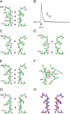
Fig. 1.
Fig. 1.
The conductive and constricted conformations of the K+ selectivity filter. (A) Close-up view of the selectivity filter of wild-type KcsA channel at high K+ concentration [K+] (PDB ID code: 1k4c). Two diagonally opposite subunits are shown in stick representation. K+ ions are shown as purple spheres. (B) Macroscopic currents of the wild-type KcsA channel elicited by a pH jump show inactivation. Currents were elicited at +100 mV by a rapid change of solution pH, at the arrow, from pH 7.5 (10 mM Hepes-KOH, 200 mM KCl) to pH 3.0 (10 mM succinate, 200 mM KCl). The selectivity filter of the KcsA channels at low [K+] (C, PDB ID code: 1k4d) and in the 32-Ǻ open structure (D, PDB ID code: 3f5w) show the constricted conformation. A rotation of the Val76–Gly77 bond causes constriction of the pore. The Gly77 Cα–Cα distance in the opposite subunits is 8.1 Å for the conductive conformation and 5.4–5.5 Å for the constricted conformation at low [K+] or in the 32-Å open state. (E) Structure of the selectivity filter of KcsAG77dA at high [K+] (PDB ID code: 2ih3). (F) A hypothetical structure of the KcsAG77dA selectivity filter in the constricted conformation. Two adjacent subunits are shown. The methyl side chain of d-Ala77 of one subunit and the carbonyl oxygen atoms of the Val76 and d-Ala77 in the adjacent subunit that clash are shown in van der Waals (VDW) representation. (G) Structure of the selectivity filter of KcsAG77dA at low [K+] (PDB ID code: 2ih1). (H) Superposition of the selectivity filter of the KcsAG77dA in high [K+] (blue) and low [K+] (red) shows that the d-Ala substitution in the selectivity filter blocks the constricted conformation.
Fig. 2.
Fig. 2.
Effect of d-Ala77 substitution on inactivation in the KcsA channel. (A) Macroscopic currents for the KcsA channels were elicited at +100 mV by a jump from pH 7.5 to pH 3.0. The initial 10 s of the decay phase following peak current was fit with a single exponential to obtain τinact. (B) Single KcsA channel currents recorded at +100 mV in steady-state conditions at pH 3.0. The KcsA WT, G77dA, and G77dA + E71A channels were obtained by semisythesis whereas the KcsA-E71A channel was obtained by recombinant expression. The KcsA channels used also contain the following amino acid substitutions: S69A, V70C (at the ligation site), Q58A, T61S, R64D (to confer AgTx2 sensitivity), and A98G (to increase the open probability of KcsA) (S2 Text, Table S1). The pH 3.0 solution is 10 mM succinate, 200 mM KCl whereas the pH 7.5 solution is 10 mM Hepes-KOH, 200 mM KCl.
Fig. 3.
Fig. 3.
Inactivation in the KvAP channel. (A) KvAP currents elicited by depolarization to +100 mV in 150 mM KCl and 10 mM Hepes-KOH (pH 7.5) show inactivation. (B) Close-up view of the selectivity filter of the KvAP channel (PDB ID code: 1orq). Two diagonally opposite subunits are shown in stick representation. K+ ions are shown as purple spheres. Residues V192 and Y199 in the KvAP channel (equivalent to V438 and Y445 in the Shaker B K+ channel) are indicated. (C) Normalized currents for the wild-type KvAP channel elicited by depolarization to +100 mV in 8 mM and 150 mM KCl with 10 mM Hepes-KOH (pH 7.5) and 150 mM RbCl with 10 mM Hepes-RbOH (pH 7.5). (D) Normalized currents for the wild-type, Y199F, and V192A KvAP channels by depolarization to +100 mV in 150 mM KCl. All of the recordings were carried out in (3:1) POPE (1-palmitoyl-2-oleoylglycero-3-phosphoethanolamine):POPG (1-palmitoyl-2-oleoylglycero-3-phosphoglycerol) lipid bilayers with a holding potential of −100 mV.
Fig. 4.
Fig. 4.
Modular semisynthesis of the KvAP channel. (A) The KvAP polypeptide is assembled by two sequential NCL reactions. In the first NCL reaction, a recombinantly expressed N-Cys peptide is ligated to a chemically synthesized filter peptide thioester to yield the intermediate peptide. The Thz protecting group (pink sphere) on the N-terminal Cys of the intermediate peptide is removed; the deprotected intermediate peptide is purified and then ligated to a recombinantly expressed thioester peptide to yield the KvAP polypeptide. The KvAP polypeptide is purified and folded in vitro to the native state. The protein segment obtained by chemical synthesis is colored red whereas the protein segments obtained by recombinant means are colored gray. The ligation sites are represented by yellow circles or spheres. (B) SDS/PAGE of the first NCL reaction between the recombinant N-Cys peptide (S6, residues 211–282) and the filter peptide (P, residues 191–210) to form the intermediate peptide (P+S6, residues 191–282) at 0 min (lane 1) and 2 h (lane 2). (C) SDS/PAGE of the second ligation reaction between the S1-5 thioester (residues 1–190) and the intermediate peptide to form the KvAP polypeptide at 0 min (lane 1) and 1 h (lane 2). (D) Size-exclusion chromatography of the semisynthetic KvAP channel. Inset shows glutaraldehyde cross-linking of the peak fraction: lane 1, without cross-linker; lane 2, cross-linked with 0.1% glutaraldehyde. The oligomeric nature (1×, 2×, and 4×) of the cross-linked bands is indicated. (E) Single-channel trace for the semisynthetic KvAP channel recorded at +100 mV. The single-channel current as a function of voltage for the semisynthetic (triangles, n = 16) and native (circles, n = 11) KvAP channels. Data are represented as mean ± SD. (F) Voltage-activated macroscopic currents from the semisynthetic KvAP channel recorded using the voltage protocol shown (Inset). (G) Voltage-dependent gating of the semisynthetic (triangles) and native (circles) KvAP channels. Tail currents were recorded after the test voltage pulse by stepping to −100mV. The fraction of the maximal current observed was plotted as a function of the test potential. The smooth line corresponds to a Boltzmann function with a V0.5 of −40 mV and a z of 1.65. The recordings shown in E and F were conducted on KvAP channels reconstituted into planar lipid bilayers composed of 1, 2-diphytanoylglycero-3-phosphocholine using symmetrical 150 mM KCl and 10 mM Hepes-KOH (pH 7.5).
Fig. 5.
Fig. 5.
Effect of d-Ala198 substitution on inactivation in the KvAP channel. (A) The single-channel current as a function of voltage for the KvAPG198dA (circles, n = 13) and the control (squares, n = 3) KvAP channels. (Inset) A single-channel trace for the KvAPG198dA channel recorded at +100 mV. The control KvAP channels contain the V191C, V192A, and V211C amino acid substitutions, and KvAPG198dA contains the G198dA substitution in addition. (B) Normalized currents for the KvAPG198dA and the control channels elicited by a depolarization to +100 mV from a holding potential of −100 mV. (C) Recovery from inactivation. Macroscopic currents for KvAPG198dA were elicited by two 5-s pulses (black first, gray second) to 100 mV, separated by holding at −100 mV for a variable duration (from 1 to 120 s). The fractional recovery was measured by the ratio of the peak current in the second pulse to the peak current in the first pulse. (D) Fractional recovery plotted as a function of the interpulse duration for KvAPG198dA (circles, n = 5) and the control channel (squares, n = 5). Solid lines represent single exponential fits used to obtain the time constant for recovery from inactivation (39.9 s for KvAPG198dA and 29.0 s for the wild-type control). The recordings shown in AC were conducted on KvAP channels reconstituted into planar lipid bilayers composed of (3:1) POPE:POPG using symmetrical 150 mM KCl and 10 mM Hepes-KOH (pH 7.5). For A and D, data are presented as mean ± SD.

References

    1. MacKinnon R. Nobel Lecture. Potassium channels and the atomic basis of selective ion conduction. Biosci Rep. 2004;24(2):75–100. - PubMed
    1. Doyle DA, et al. The structure of the potassium channel: Molecular basis of K+ conduction and selectivity. Science. 1998;280(5360):69–77. - PubMed
    1. Zhou Y, Morais-Cabral JH, Kaufman A, MacKinnon R. Chemistry of ion coordination and hydration revealed by a K+ channel-Fab complex at 2.0 A resolution. Nature. 2001;414(6859):43–48. - PubMed
    1. McCoy JG, Nimigean CM. Structural correlates of selectivity and inactivation in potassium channels. Biochim Biophys Acta. 2012;1818(2):272–285. - PMC - PubMed
    1. Hoshi T, Zagotta WN, Aldrich RW. Two types of inactivation in Shaker K+ channels: Effects of alterations in the carboxy-terminal region. Neuron. 1991;7(4):547–556. - PubMed

Publication types

LinkOut - more resources