. 2013 Sep 24;110(39):15716-21.
doi: 10.1073/pnas.1305420110. Epub 2013 Sep 9.

Dissection of Cdk1-cyclin complexes in vivo

Affiliations

Dissection of Cdk1-cyclin complexes in vivo

Po Hien Ear et al. Proc Natl Acad Sci U S A. .

Abstract

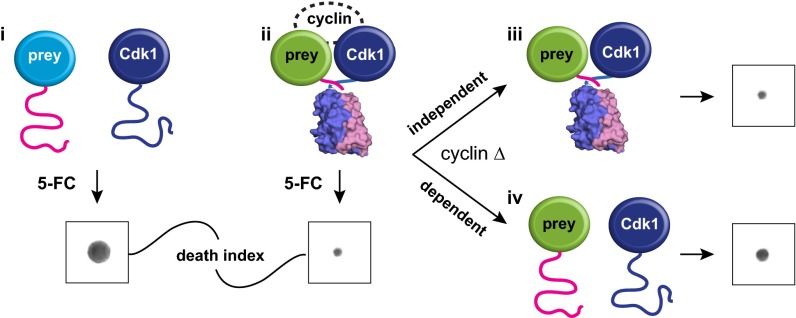
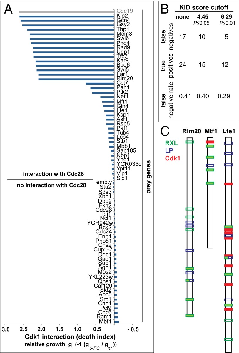
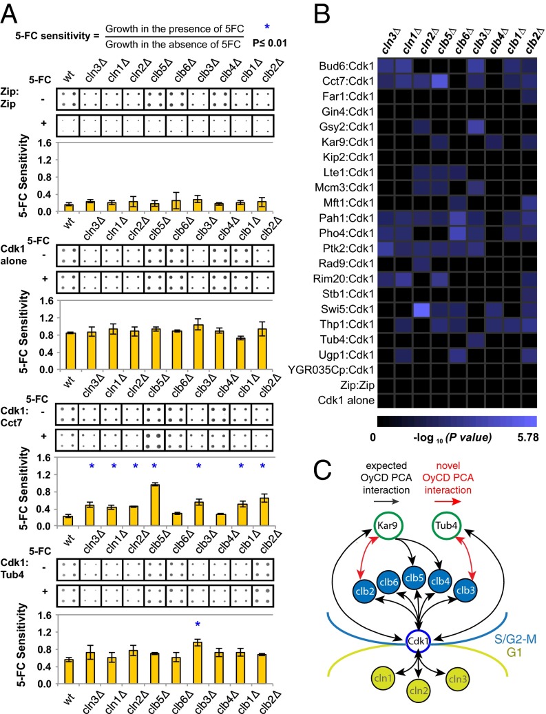
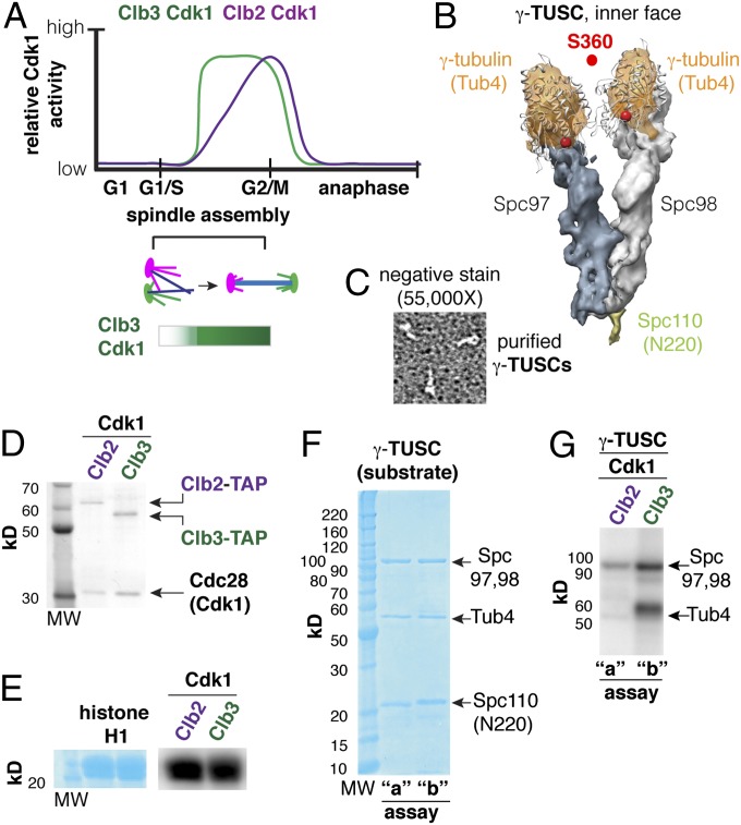
Cyclin-dependent kinases (Cdks) are regulatory enzymes with temporal and spatial selectivity for their protein substrates that are governed by cell cycle-regulated cyclin subunits. Specific cyclin-Cdk complexes bind to and phosphorylate target proteins, coupling their activity to cell cycle states. The identification of specific cyclin-Cdk substrates is challenging and so far, has largely been achieved through indirect correlation or use of in vitro techniques. Here, we use a protein-fragment complementation assay based on the optimized yeast cytosine deaminase to systematically identify candidate substrates of budding yeast Saccharomyces cerevisiae Cdk1 and show dependency on one or more regulatory cyclins. We identified known and candidate cyclin dependencies for many predicted protein kinase Cdk1 targets and showed elusory Clb3-Cdk1-specific phosphorylation of γ-tubulin, thus establishing the timing of this event in controlling assembly of the mitotic spindle. Our strategy can be generally applied to identify substrates and accessory subunits of multisubunit protein complexes.

Keywords: cyclin specificity; in vivo enzyme complexes screen.


Conflict of interest statement

The authors declare no conflict of interest.

Figures

Fig. 1.
Fig. 1.
Dissecting Cdk1 complexes using the OyCD PCA. Detecting the interaction between Cdk1 and a protein of interest using the OyCD PCA. Cdk1 and proteins of interest (prey proteins) are fused to OyCD fragments. In the death selection OyCD PCA, (i) the absence of an interaction fails to allow the OyCD reporter enzyme to fold and restore the activity of the native enzyme. Cells expressing these fusion proteins are resistant to 5-FC. (ii) If prey protein interacts with Cdk1, cells are sensitive to 5-FC. When a cyclin gene is deleted (cyclin Δ), (iii) a prey protein can still interact with Cdk1, allowing the reporter fragments to fold; consequently, cells are sensitive to 5-FC. The prey protein–Cdk1 interaction is independent of any cyclin. (iv) If the interaction is cyclin-dependent, the absence of a specific cyclin results in partial or total resistance to 5-FC.
Fig. 2.
Fig. 2.
Identification of interaction partners of Cdk1. (A) Quantification of Cdk1–prey protein interactions. Death index is calculated as the log10 of the ratio of pixel mean intensities of 5-FC–treated divided by untreated colonies (5-FC/not treated). The Cdk1–Cdc19 interaction was found to be a technical false positive (see text). (B) Estimation of the FNR for 37 proteins identified to interact with Cdk1 by OyCD PCA (details of calculation in SI Text). (C) Examples of Cdk1 interacting partners with cyclin binding motifs and with (Lte1 and Mft1) or without (Rim20) consensus Cdk1 phosphorylation sites. Filled bars are high-quality sites (P ≤ 0.01, open bars are low-quality sites).
Fig. 3.
Fig. 3.
Cyclin dependence of Cdk1–prey protein interactions. (A) Quantification of cyclin dependence. The overexpression of some fusion genes slightly affects the fitness of the yeast strain compared with yeast expressing only Cdk1 or Zip:Zip fused to OyCD fragments. The deletion of some cyclin genes affects the ability of the strain to grow compared with the WT strain. The effect of the OyCD PCA activity on growth is relatively constant in the different cyclin deletion strains (mean ± SD). Statistical significance was assessed using Student t test. P = 0.04 was obtained for WT and cln3Δ strains expressing Zip:Zip interaction. P = 0.02 was obtained for WT and clb1Δ deletion strains expressing Cdk1 alone (contingency of the Cdk1 complexes). The gene of interest and Cdk1 fused to OyCD fragments were transformed into the FCY1Δ deletion strain, which is referred to as the WT, as well as in nine different cyclins and FCY1Δ double deletion strains, which are represented by their gene name in italic followed by a Δ (e.g., cln1Δ). Four colonies of each transformation were assayed for OyCD PCA activity in the presence of 1 mg/mL 5-FC in three different experiments. The growth of each sample was quantified using ImageJ. Only the results of one set of a quadruple experiment are represented. All strains expressing only Cdk1–OyCD-F[2] (Cdk1 alone) are resistant to the 5-FC death selection assay with P < 0.02. All strains expressing the GCN4 leucine zipper domains (Zip:Zip) fused to the OyCD fragments are sensitive to 5-FC in the death selection assay with a P < 0.04. A loss of interaction detected in the different cyclin deletion strains is depicted with corresponding P value. P ≤ 0.01 was used as a cutoff for this experiment (indicated with blue asterisk). (B) Results for P ≤ 0.01 are represented on the matrix (44). (C) Summary of previously described (black arrows) and discovered (red arrows) interactions between Cdk1–cylin complexes, Kar9, and Tub4 and timing of their functions in the mitotic cell cycle.
Fig. 4.
Fig. 4.
γ-Tubulin is a Clb3–Cdk1 substrate in vitro. (A) Early and late B-type cyclin–Cdk complexes (e.g., Clb3 and Clb2) are active during spindle assembly, with Clb3 activity peaking before Clb2 reaches its maximum activity. (B) The γ-tubulin small complex (γ-TUSC) is composed of Tub4, Spc97, and Spc98 (2:1:1 stoichiometry). γ-TUSCs are Y-shaped complexes that form spontaneously in coinfected Sf9 cells in the presence of a fragment of Spc110 (N-terminal 220). (C) Verification of the structure of purified γ-TUSCs using negative staining and transmission electron microscopy (TEM). (D) Clb–Cdk1 complexes purified from yeast. (E) Histone H1 was used as a control to verify Clb–Cdk1 activity. (F) γ-TUSC substrate (0.4 µM) reacted with Clb–Cdk1 in parallel kinase assays: a, 2 nM Clb2–Cdk1; b, 5 nM Clb3–Cdk1 for 30 min at 30 °C. (G) Tub4 was efficiently phosphorylated by Clb3–Cdk1 but not Clb2–Cdk1, whereas Spc97 and -98 are phosphorylated by both Clb2–Cdk1 and Clb3–Cdk1.

References

    1. Thornton BR, Toczyski DP. Precise destruction: An emerging picture of the APC. Genes Dev. 2006;20(22):3069–3078. - PubMed
    1. Cohen P. The structure and regulation of protein phosphatases. Annu Rev Biochem. 1989;58(1989):453–508. - PubMed
    1. Morgan DO. Cyclin-dependent kinases: Engines, clocks, and microprocessors. Annu Rev Cell Dev Biol. 1997;13(1997):261–291. - PubMed
    1. Janssens V, Longin S, Goris J. PP2A holoenzyme assembly: In cauda venenum (the sting is in the tail) Trends Biochem Sci. 2008;33(3):113–121. - PubMed
    1. Morgan DO. Principles of CDK regulation. Nature. 1995;374(6518):131–134. - PubMed

Publication types

MeSH terms