. 2013 Sep 17;110(38):E3595-604.
doi: 10.1073/pnas.1220050110. Epub 2013 Sep 9.

Mechanism for activation of mutated epidermal growth factor receptors in lung cancer

Affiliations

Mechanism for activation of mutated epidermal growth factor receptors in lung cancer

Monica Red Brewer et al. Proc Natl Acad Sci U S A. .

Erratum in

  • Proc Natl Acad Sci U S A. 2013 Dec 10;110(50):20344

Abstract

The initiation of epidermal growth factor receptor (EGFR) kinase activity proceeds via an asymmetric dimerization mechanism in which a "donor" tyrosine kinase domain (TKD) contacts an "acceptor" TKD, leading to its activation. In the context of a ligand-induced dimer, identical wild-type EGFR TKDs are thought to assume the donor or acceptor roles in a random manner. Here, we present biochemical reconstitution data demonstrating that activated EGFR mutants found in lung cancer preferentially assume the acceptor role when coexpressed with WT EGFR. Mutated EGFRs show enhanced association with WT EGFR, leading to hyperphosphorylation of the WT counterpart. Mutated EGFRs also hyperphosphorylate the related erythroblastic leukemia viral oncogene (ErbB) family member, ErbB-2, in a similar manner. This directional "superacceptor activity" is particularly pronounced in the drug-resistant L834R/T766M mutant. A 4-Å crystal structure of this mutant in the active conformation reveals an asymmetric dimer interface that is essentially the same as that in WT EGFR. Asymmetric dimer formation induces an allosteric conformational change in the acceptor subunit. Thus, superacceptor activity likely arises simply from a lower energetic cost associated with this conformational change in the mutant EGFR compared with WT, rather than from any structural alteration that impairs the donor role of the mutant. Collectively, these findings define a previously unrecognized mode of mutant-specific intermolecular regulation for ErbB receptors, knowledge of which could potentially be exploited for therapeutic benefit.

Keywords: TKI; WZ-4002; lapatinib; mutation.


Conflict of interest statement

Conflict of interest statement: Rights to testing of EGFR T790M mutations were licensed by Memorial Sloan-Kettering Cancer Center on behalf of W.P. and others to MolecularMD.

Figures

Fig. 1.
Fig. 1.
Allosteric activation of the WT EGFR kinase. Kinase activation occurs by asymmetric dimerization of tyrosine kinase domains (TKDs), creating the asymmetric dimerization interface (Far Left). Mutations at specific residues in this interface enforce TKDs to act solely as donors (I682Q) or acceptors (V924R), which are inactive when expressed alone due to an inability to dimerize (Center two panels). Coexpression of an enforced donor with an enforced acceptor restores dimerization capacity to 50% wild-type levels (Far Right).
Fig. 2.
Fig. 2.
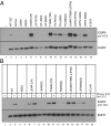
N-lobe dimerization interface mutations have different effects in WT and lung-cancer–associated EGFRs. EGFR-ICD constructs (A) or full-length EGFR constructs (B) encoding either WT or kinase domain mutants (LR, L834R; TM, T766M; LRTM, L834R/T766M) with or without N-lobe mutations at the asymmetric dimer interface were transiently expressed in HEK293 cells. In B, cells were serum starved and treated with or without 50 ng/mL EGF for 10 min as indicated. Lysates were subjected to SDS/PAGE and immunoblotting for EGFR-pY1173 and total EGFR.
Fig. 3.
Fig. 3.
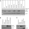
C-lobe dimerization interface mutations have different effects in WT and lung-cancer–associated EGFRs and ErbB-4. (A) Full-length EGFR constructs encoding either WT or kinase domain mutants (LR, L834R; TM, T766M; LRTM, L834R/T766M) with or without C-lobe mutations at the asymmetric dimer interface were transiently expressed in HEK293 cells. Cells were serum starved and treated with or without 50 ng/mL EGF for 10 min as indicated. Lysates were subjected to SDS/PAGE and immunoblotting for EGFR-pY1173 and total EGFR. (B) EGFR-ICD constructs encoding either WT or kinase domain mutants (LRTM, L834R/T766M) with or without single or double C-lobe mutations at the asymmetric dimer interface were transiently expressed in HEK293 cells. Lysates were subjected to SDS/PAGE and immunoblotting for EGFR-pY1173 and total EGFR. (C) ErbB-4–ICD constructs encoding either WT or kinase domain mutant (T796M) with or without C-lobe mutation at the asymmetric dimer interface were transiently expressed in HEK293 cells. Lysates were subjected to SDS/PAGE and immunoblotting for anti-pY and total ErbB-4. Two times total lysate was loaded in lanes labeled as 2×.
Fig. 4.
Fig. 4.
Lung-cancer–associated EGFRs demonstrate altered donor/acceptor biochemical complementation properties but still form asymmetric dimers. (A) ICD constructs containing mutations that enforce either donor-only (I682Q) or acceptor-only (V924R) behavior in the WT asymmetric dimer were used to assess the mechanism of activation of the lung-cancer–associated mutant L834R/T766M EGFR (LRTM). The indicated combinations of ICD constructs were coexpressed in HEK293 cells, and lysates were subjected to SDS/PAGE and immunoblotting. As predicted by the allosteric model of activation, autophosphorylation levels of coexpressed WT V924R and WT I682Q (lane 5) are half that of WT EGFR (lane 2). Autophosphorylation levels of coexpressed LRTM/I682Q and LRTM/V924R are not greater than either ICD expressed alone (lane 9 compared with lanes 7 and 8), showing that the mutated ICD behaves differently from WT. Coexpression of a WT acceptor mutant (V924R) with LRTM/I682Q dramatically diminishes activity (lane 10). By contrast, coexpression of LRTM/V924R with a WT donor mutant (I682Q) results in apparent superacceptor activity as described in the text (lane 11). (B) Crystal structure of L834R/T766M reveals an asymmetric dimer. The crystals contain two independent molecules that form a typical asymmetric dimer interaction. The L834R and T766M mutations are indicated, as are the locations of I682 and V924 in the asymmetric dimer interface. The irreversible inhibitor PD168393 was included to stabilize the protein and facilitate crystallization. See Fig. S5 for a comparison of the asymmetric dimer interface in this structure with that of WT EGFR.
Fig. 5.
Fig. 5.
Lung-cancer–associated mutated EGFR acceptor activity is concomitant with increased WT EGFR monomer association and hyperphosphorylation. (A) Coexpression of ICD constructs containing the donor-enforcing I682Q in a WT background (green) or the acceptor-enforcing V924R (blue) in the lung-cancer–associated mutant backgrounds L834R (LR) or L834R/T766M EGFR (LRTM) was used to assess the level of monomer association. Lysates from the indicated combinations were subjected to immunoprecipitation with anti-Flag, followed by SDS/PAGE and immunoblotting for protein levels with anti-Myc and anti-Flag as indicated. Levels of coimmunoprecipitated acceptor-enforced mutant ICDs with donor I682Q are greater than levels of coimmunoprecipitated levels of V924R with I682Q (lanes 4 and 5 compared with lane 3), indicating that the lung-cancer–associated mutations increase the affinity of these interactions. (B) Coexpression of increasing amounts of Y992 truncated LRTM-ICD relative to a fixed amount of Flag-tagged WT ICD results in hyperphosphorylation of the WT population as assessed by Western blotting for anti-pY1173 and anti-EGFR. (C) Coexpression of increasing amounts (as indicated in micrograms) of myc-tagged lung-cancer–associated mutant ICDs relative to a fixed amount of Flag-tagged WT ICD results in dose-dependent hyperphosphorylation of the WT population as assessed by Flag immunoprecipitation and subsequent Western blotting for antiphosphotyrosine. Lysates were also immunoblotted with anti–EGFR-pY1173, anti-Myc, and anti-Flag, which show that the level of pY1173 signal cannot be explained solely by the level of the Myc-tagged ICDs harboring lung-cancer–associated mutations. Also, more L834R-ICD (LR) compared with L834R/T766M-ICD (LRTM) is required to hyperphosphorylate WT ICD to levels equivalent to that of the Flag-tagged TKD mutant ICD (compare lanes 4 with 6 and 9 with 11).
Fig. 6.
Fig. 6.
Hyperphosphorylation of WT-EGFR-ICD in the presence of lung-cancer–mutated EGFR-ICD is insensitive to lapatinib, whereas WZ-4002 restores normal WT EGFR-ICD tyrosine phosphorylation levels. (A) Coexpression of myc-tagged lung-cancer–associated mutant ICD with 1.0 μg of Flag-tagged WT ICD results in hyperphosphorylation of the WT population as assessed by anti-Flag immunoprecipitation and subsequent Western blotting for antiphosphotyrosine. Lapatinib treatment (100 nM) results in inhibition of kinase activity of singly expressed WT EGFR-ICD, but not inhibition of the hyperphosphorylation induced by coexpressing 1/10th the concentration of LRTM-myc-ICD, consistent with trans-phosphorylation by the lapatinib-insensitive mutated TKDs. (B) Coexpression of myc-tagged lung-cancer–associated mutant ICD with 1.0 μg of Flag-tagged WT ICD results in hyperphosphorylation of the WT population as assessed by anti-Flag immunoprecipitation and subsequent Western blotting for antiphosphotyrosine. Treatment of WT- and LRTM-ICD coexpressing cells with the T766M-specific TKI WZ-4002 (100 nM), reduces hyperphosphorylation of the WT ICD (lane 5). WZ-4002 treatment does not inhibit WT kinase activity of singly expressed WT-EGFR-ICD, but conversely, completely inhibits kinase activity of singly expressed LRTM-ICD-myc as assessed by Western blotting (lanes 3 and 7, respectively).
Fig. 7.
Fig. 7.
Lung-cancer–associated EGFRs induce hyperphosphorylation of coexpressed ErbB-2 by virtue of superacceptor activity. (A) Coexpression of lung-cancer–associated mutant ICDs with WT ErbB-2 ICDs in HEK293 cells results in hyperphosphorylation of the ErbB-2–ICD population as assessed by anti–ErbB-2 immunoprecipitation and subsequent Western blotting for anti–ErbB-2–pY1221/22. Coexpression of WT EGFR-ICD with ErbB-2–ICD results in a slight increase in phospho–ErbB-2, whereas coexpression of either lung-cancer–associated ICD (LR or LRTM) with ErbB-2 results in hyperphosphorylation of ErbB-2–ICD (lanes 4, 8, and 12, respectively). Unlike lung-cancer–associated EGFRs containing the donor-enforcing I682Q mutation, combination of lung-cancer–associated EGFR-ICDs containing acceptor-enforcing V924R mutation with ErbB-2–ICD results in hyperphosphorylation of ErbB-2–ICD at levels equivalent to the combination of ErbB-2–ICD with lung-cancer–associated parent EGFR-ICDs (lane 10 compared with lane 8 and lane 14 compared with lane 12). (B) Coexpression of intact WT EGFR with intact ErbB-2 results in an increase in tyrosine-phosphorylated ErbB-2 relative to ErbB-2 expressed alone, whereas ErbB-2 coexpression with lung-cancer–associated mutant EGFR (LRTM) results in a significant increase in phospho-ErbB-2 levels. Treatment of cells coexpressing ErbB-2 and EGFR-L834R/T766M with WZ-4002 in the presence of EGF abrogates the EGFR mutant-induced hyperphosphorylation of ErbB-2 (compare lane 16 to lane 11). A comparatively less dramatic effect on ErbB-2 tyrosine phosphorylation is observed for cells coexpressing ErbB-2 and WT EGFR treated with WZ-4002 in the presence of EGF (compare lane 15 to lane 9). (C) Coexpression of of 0.1 μg myc-tagged lung cancer-associated mutant ICD with 1.0 μg of Flag-tagged WT EGFR or ErbB-2–ICD results in additive increases in pAkt or pStat-3 levels compared with singly expressed ICDs, whereas a synergistic increase in pErk levels is observed upon coexpression (compare lanes 2–4 to lanes 6 and 7).

Comment in

  • EGFR lung cancer mutants get specialized.
    Littlefield P, Jura N. Littlefield P, et al. Proc Natl Acad Sci U S A. 2013 Sep 17;110(38):15169-70. doi: 10.1073/pnas.1314719110. Epub 2013 Sep 10. Proc Natl Acad Sci U S A. 2013. PMID: 24023066 Free PMC article. No abstract available.

References

    1. Pao W, Chmielecki J. Rational, biologically based treatment of EGFR-mutant non-small-cell lung cancer. Nat Rev Cancer. 2010;10(11):760–774. - PMC - PubMed
    1. Sharma SV, Bell DW, Settleman J, Haber DA. Epidermal growth factor receptor mutations in lung cancer. Nat Rev Cancer. 2007;7(3):169–181. - PubMed
    1. Mitsudomi T, et al. West Japan Oncology Group Gefitinib versus cisplatin plus docetaxel in patients with non-small-cell lung cancer harbouring mutations of the epidermal growth factor receptor (WJTOG3405): An open label, randomised phase 3 trial. Lancet Oncol. 2010;11(2):121–128. - PubMed
    1. Mok TS, et al. Gefitinib or carboplatin-paclitaxel in pulmonary adenocarcinoma. N Engl J Med. 2009;361(10):947–957. - PubMed
    1. Rosell R, Viteri S, Molina MA, Benlloch S, Taron M. Epidermal growth factor receptor tyrosine kinase inhibitors as first-line treatment in advanced nonsmall-cell lung cancer. Curr Opin Oncol. 2010;22(2):112–120. - PubMed

Publication types

Associated data