. 2013 Oct 25;288(43):31268-79.
doi: 10.1074/jbc.M113.462341. Epub 2013 Sep 9.

Toll-like receptor 3-mediated necrosis via TRIF, RIP3, and MLKL

Affiliations

Toll-like receptor 3-mediated necrosis via TRIF, RIP3, and MLKL

William J Kaiser et al. J Biol Chem. .

Abstract

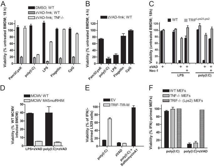
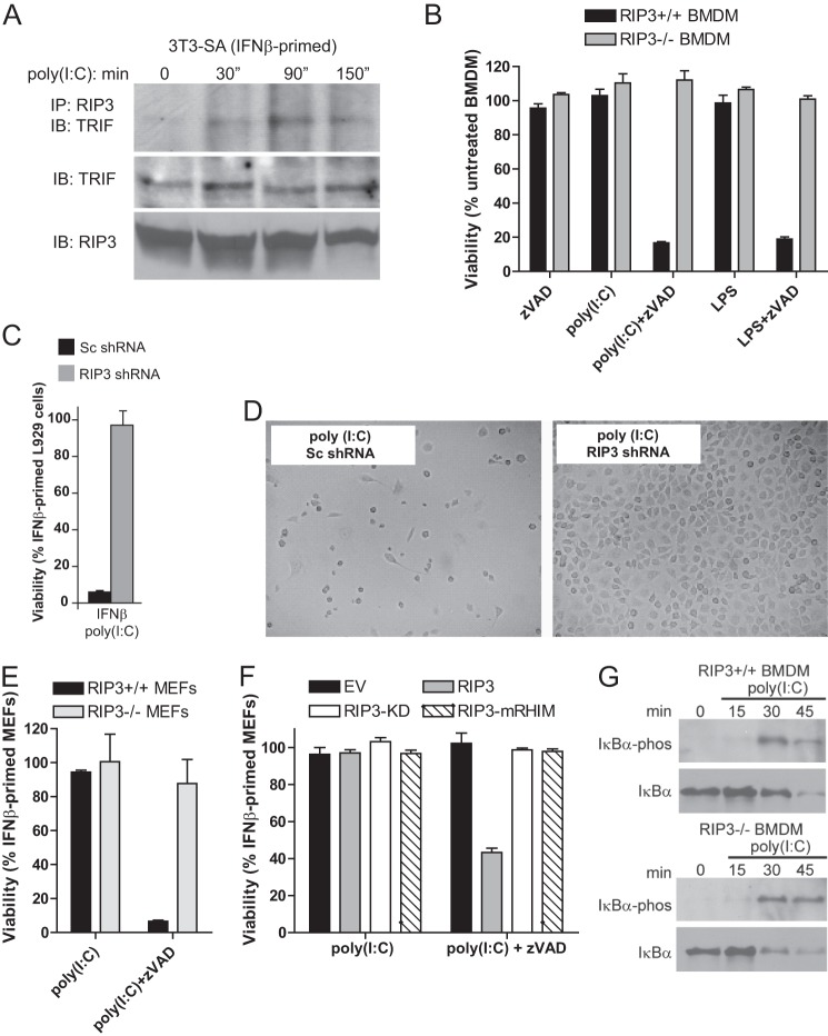
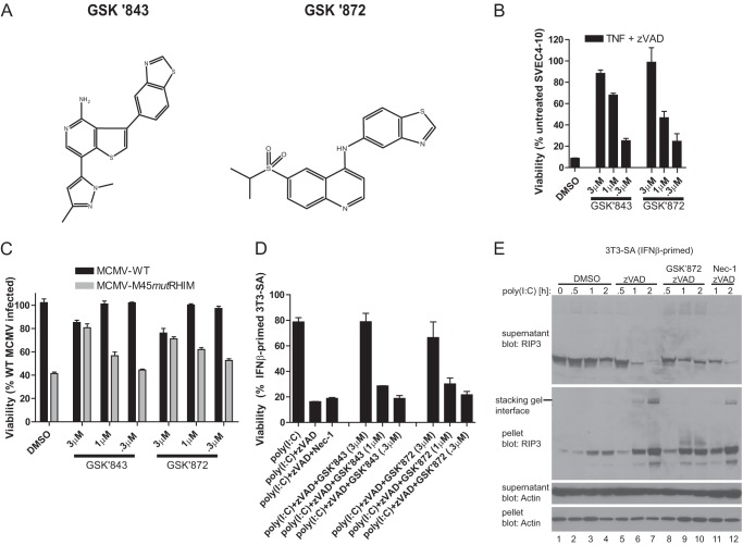
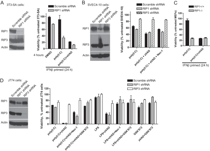
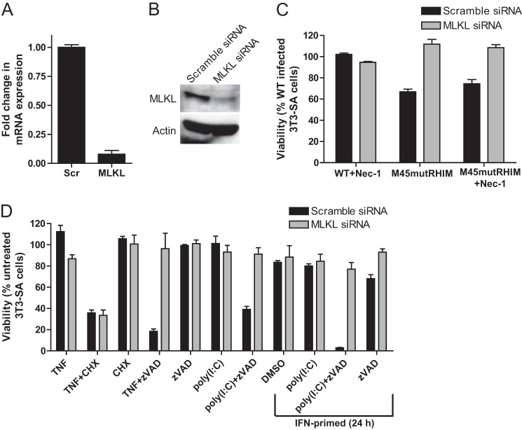
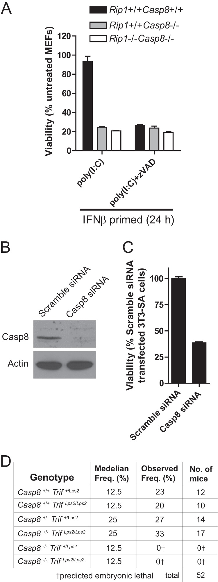
Toll-like receptor (TLR) signaling is triggered by pathogen-associated molecular patterns that mediate well established cytokine-driven pathways, activating NF-κB together with IRF3/IRF7. In addition, TLR3 drives caspase 8-regulated programmed cell death pathways reminiscent of TNF family death receptor signaling. We find that inhibition or elimination of caspase 8 during stimulation of TLR2, TLR3, TLR4, TLR5, or TLR9 results in receptor interacting protein (RIP) 3 kinase-dependent programmed necrosis that occurs through either TIR domain-containing adapter-inducing interferon-β (TRIF) or MyD88 signal transduction. TLR3 or TLR4 directly activates programmed necrosis through a RIP homotypic interaction motif-dependent association of TRIF with RIP3 kinase (also called RIPK3). In fibroblasts, this pathway proceeds independent of RIP1 or its kinase activity, but it remains dependent on mixed lineage kinase domain-like protein (MLKL) downstream of RIP3 kinase. Here, we describe two small molecule RIP3 kinase inhibitors and employ them to demonstrate the common requirement for RIP3 kinase in programmed necrosis induced by RIP1-RIP3, DAI-RIP3, and TRIF-RIP3 complexes. Cell fate decisions following TLR signaling parallel death receptor signaling and rely on caspase 8 to suppress RIP3-dependent programmed necrosis whether initiated directly by a TRIF-RIP3-MLKL pathway or indirectly via TNF activation and the RIP1-RIP3-MLKL necroptosis pathway.

Keywords: Apoptosis; Caspase; Necrosis (Necrotic Death); RIPK3; Serine/Threonine Protein Kinase; Toll-like Receptors (TLR).


Figures

FIGURE 1.
FIGURE 1.
TLR stimulation in the presence of caspase inhibitor triggers cell death. A, viability of WT and TNF−/− BMDM at 18 h after stimulation with Pam3CysK (1 μg/ml), poly(I:C) (25 μg/ml), LPS (500 ng/ml), flagellin (500 ng/ml), or CpG (1 μg/ml) in the presence of Z-VAD-fmk (25 μm) or vehicle (DMSO) control. B, viability of WT BMDM at 6 h after stimulation with the indicated TLR ligands in the presence of Z-VAD-fmk. C, viability of WT or TRIF mutant (Lps2/Lps2) BMDM at 18 h after stimulation with poly(I:C) or LPS in the presence or absence of Z-VAD-fmk. D, CellTiter-Glo assay was used to assess viability of BMDM after infection with either WT or M45mutRHIM MCMV (multiplicity of infection of 5) for 18 h followed by treatment with either LPS or poly(I:C) in the presence of Z-VAD-fmk. E, viability of IFNβ-primed L929 cells stably expressing a dominant negative TRIF-TIR domain-only construct (TRIF-TIR-M) or vector only control (EV). Cells were first primed with IFNβ (50 units/ml) for 24 h and then stimulated with poly(I:C) in the absence or presence of Z-VAD-fmk or with poly(I:C) and bafilomycin A1 (500 nm) for 18 h, as indicated. F, viability of WT, Tnf−/−, or TrifLps2/Lps2 MEFs at 18 h after stimulation with TLR3 agonist poly(I:C) in the presence of Z-VAD-fmk. Cell viability was assessed by determining ATP levels (CellTiter-Glo, Promega). Error bars, S.D.
FIGURE 2.
FIGURE 2.
TLR3-induced programmed necrosis requires RIP3. A, following priming of 3T3-SA cells for 24 h, cells were stimulated with poly(I:C) (25 μg/ml). RIP3 was immunoprecipitated (IP) from cell lysates to detect an interaction with TRIF. IB, immunoblot. B–F, viability assays at 18 h after stimulation with poly(I:C) in the presence of Z-VAD-fmk, showing WT (Rip3+/+) or Rip3−/− BMDM (B); IFNβ-primed L929 cells expressing control scramble (Sc) or RIP3-specific shRNA (C); photomicrographs of IFNβ-primed L929 cells expressing control scramble or RIP3-specific shRNA (D); IFNβ-primed WT (Rip3+/+) or Rip3−/− MEFs (E); Rip3−/− MEFs reconstituted with WT RIP3, RIP3-KD, or RIP3-mRHIM (F). Cell viability in B–F was determined by ATP assay. G, immunoblot detection of total and phosphorylated IkBα in WT (Rip3+/+, left panel) or Rip3−/− (right panel) BMDM following stimulation with poly(I:C) for the indicated times (minutes).
FIGURE 3.
FIGURE 3.
Role of RIP3 kinase in TLR3-induced programmed necrosis. A, chemical structure of compounds GSK'843 and GSK'872. B, viability of 3T3-SA cells at 18 h after treatment with TNF in the presence of Z-VAD-fmk in vehicle control (DMSO) or treated with the indicated concentrations of RIP3 kinase inhibitors, GSK'843 or GSK'872. C, viability of SVEC4-10 cells at 18 h post-infection with WT or M45mutRHIM MCMV in vehicle control (DMSO) or treated with the indicated concentrations of RIP3 kinase inhibitors. D, viability of IFNβ-primed 3T3-SA cells at 18 h after stimulation with poly(I:C) in the absence or presence of Z-VAD-fmk and treatment with Nec-1 (30 μm) or the indicated concentrations of RIP3 kinase inhibitors. E, immunoblot detecting RIP3 and β-actin present in the soluble (supernatant) and insoluble (pellet) fraction following stimulation of 3T3-SA cells with poly(I:C) for the indicated times (hours) in the absence or presence of the caspase inhibitor Z-VAD-fmk and GSK'872 (3 μm) or Nec-1 (30 μm). Cell viability was determined by the ATP assay. Inquiries about RIP3 kinase inhibitors GSK'843 and GSK'872 should be directed to P. Gough (peter.j.gough@gsk.com).
FIGURE 4.
FIGURE 4.
Differential role of RIP1 in TLR-induced necrosis in macrophages versus other cell types. A, viability of IFNβ-primed 3T3-SA cells transfected with either RIP1 or MLKL siRNA smartpools. Cells were stimulated with poly(I:C) in the absence or presence of Z-VAD for 4 h. B, viability of SVEC4-10 cells expressing control scramble and RIP1-specific or RIP3-specific shRNA in the absence or presence of Z-VAD-fmk and Nec-1 (30 μm) for 18 h. C, WT (Rip1+/+) or Rip1−/− MEFs at 18 h after stimulation with poly(I:C) in the absence or presence of Z-VAD-fmk and IFNβ. D, J774 macrophages after 18 h of stimulation with LPS or poly(I:C) in the absence or presence of Z-VAD-fmk, Nec-1, and GSK'872. Cell viability was determined by the ATP assay.
FIGURE 5.
FIGURE 5.
Role of MLKL in TLR3- and DAI-induced necrosis. 3T3-SA cells were transfected with either MLKL or scramble (Scr) siRNA pools. A, at 48 h post-transfection, quantitative real time PCR detected the fold change in MLKL mRNA relative to β-actin. B, immunoblot analysis of MLKL and β-actin in siRNA-transfected 3T3-SA cell. C, viability of 3T3-SA cells at 18 h post-infection with WT or M45mutRHIM MCMV. Cells were infected in the presence of vehicle control (DMSO) or 30 μm Nec-1. D, viability of siRNA-transfected 3T3-SA cells at 18 h after stimulation with TNF or poly(I:C) in the absence or presence of Z-VAD-fmk or cycloheximide (CHX). Cells were primed with IFNβ for 24 prior to stimulation where indicated. Cell viability was determined by the ATP assay.
FIGURE 6.
FIGURE 6.
Casp8 suppression of TLR3-mediated TRIF- and RIP3-dependent programmed necrosis. A, viability of WT, Casp8−/−, or Casp8−/−Rip1−/− MEFs at 18 h after stimulation with poly(I:C) in the absence or presence of Z-VAD-fmk. B, 3T3-SA cells were transfected with either the Casp8 or Scramble siRNA pools. At 72 h post-transfection Casp8 and β-actin levels were determined by immunoblot analysis. C, cell viability was determined. A and C, cell viability was determined by ATP levels. Error bars, S.D. D, epistatic analysis of mice born following a Casp8+/−Trif+/Lps2 × Casp8+/−TrifLps2/Lps2 intercross with predicted and observed frequencies.

References

    1. Lamkanfi M., Dixit V. M. (2010) Manipulation of host cell death pathways during microbial infections. Cell Host Microbe 8, 44–54 - PubMed
    1. Everett H., McFadden G. (1999) Apoptosis: an innate immune response to virus infection. Trends Microbiol. 7, 160–165 - PubMed
    1. Kumar H., Kawai T., Akira S. (2011) Pathogen recognition by the innate immune system. Int. Rev. Immunol. 30, 16–34 - PubMed
    1. Kaiser W. J., Offermann M. K. (2005) Apoptosis induced by the toll-like receptor adapter TRIF is dependent on its receptor interacting protein homotypic interaction motif. J. Immunol. 174, 4942–4952 - PubMed
    1. He S., Liang Y., Shao F., Wang X. (2011) Toll-like receptors activate programmed necrosis in macrophages through a receptor-interacting kinase-3-mediated pathway. Proc. Natl. Acad. Sci. U.S.A. 108, 20054–20059 - PMC - PubMed

Publication types

MeSH terms