

MRI findings of elevated intracranial pressure in cerebral venous thrombosis versus idiopathic intracranial hypertension with transverse sinus stenosis
- PMID: 24019557
- PMCID: PMC3765015
- DOI: 10.3109/01658107.2012.738759
MRI findings of elevated intracranial pressure in cerebral venous thrombosis versus idiopathic intracranial hypertension with transverse sinus stenosis
Abstract
Purpose: To determine whether MRI signs suggesting elevated intracranial pressure (ICP) are preferentially found in patients with idiopathic intracranial hypertension (IIH) than in those with cerebral venous thrombosis (CVT).
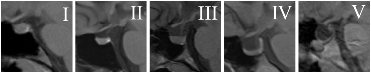
Methods: Among 240 patients who underwent standardized contrast-enhanced brain MRI/MRV at our institution between 9/2009 and 9/2011, 60 with abnormal imaging findings on MRV were included: 27 patients with definite IIH, 2 patients with presumed IIH, and 31 with definite CVT. Medical records were reviewed, and imaging studies were prospectively evaluated by the same neuroradiologist to assess for presence or absence of transverse sinus stenosis (TSS), site of CVT if present, posterior globe flattening, optic nerve sheath dilation/tortuosity, and the size/appearance of the sella turcica.
Results: 29 IIH patients (28 women, 19 black, median-age 28, median-body mass index, 34) had bilateral TSS. 31 CVT patients (19 women, 13 black, median-age 46, median-BMI 29) had thrombosis of the sagittal (3), sigmoid (3), cavernous (1), unilateral transverse (7), or multiple (16) sinuses or cortical veins (1). Empty/partially-empty sellae were more common in IIH (3/29 and 24/29) than in CVT patients (1/31 and 19/31) (p<0.001). Flattening of the globes and dilation/tortuosity of the optic nerve sheaths were more common in IIH (20/29 and 18/29) than in CVT patients (13/31 and 5/31) (p<0.04).
Conclusion: Although abnormal imaging findings suggestive of raised ICP are more common in IIH, they are not specific for IIH and are found in patients with raised ICP from other causes such as CVT.
Keywords: MRI; MRV; cerebral venous thrombosis; empty sella; idiopathic intracranial hypertension; venous hypertension.
Figures


Similar articles
-
Does bilateral transverse cerebral venous sinus stenosis exist in patients without increased intracranial pressure?Clin Neurol Neurosurg. 2013 Aug;115(8):1215-9. doi: 10.1016/j.clineuro.2012.11.004. Epub 2012 Dec 5. Clin Neurol Neurosurg. 2013. PMID: 23219404 Free PMC article.
-
Imaging signs in idiopathic intracranial hypertension: Are these signs seen in secondary intracranial hypertension too?Ann Indian Acad Neurol. 2013 Apr;16(2):229-33. doi: 10.4103/0972-2327.112476. Ann Indian Acad Neurol. 2013. PMID: 23956571 Free PMC article.
-
Impact of rater experience and referral question on detecting magnetic resonance imaging features of idiopathic intracranial hypertension.Eur J Neurol. 2023 Oct;30(10):3314-3321. doi: 10.1111/ene.15986. Epub 2023 Jul 27. Eur J Neurol. 2023. PMID: 37475659
-
MRI findings as markers of idiopathic intracranial hypertension.Curr Opin Neurol. 2021 Feb 1;34(1):75-83. doi: 10.1097/WCO.0000000000000885. Curr Opin Neurol. 2021. PMID: 33230036 Free PMC article. Review.
-
Brain Imaging in Idiopathic Intracranial Hypertension.J Neuroophthalmol. 2015 Dec;35(4):400-11. doi: 10.1097/WNO.0000000000000303. J Neuroophthalmol. 2015. PMID: 26457687 Review.
Cited by
-
Empty Sella Is a Sign of Symptomatic Lateral Sinus Stenosis and Not Intracranial Hypertension.AJNR Am J Neuroradiol. 2019 Oct;40(10):1695-1700. doi: 10.3174/ajnr.A6210. Epub 2019 Sep 19. AJNR Am J Neuroradiol. 2019. PMID: 31537518 Free PMC article.
-
The comparative analysis of non-thrombotic internal jugular vein stenosis and cerebral venous sinus stenosis.J Thromb Thrombolysis. 2019 Jul;48(1):61-67. doi: 10.1007/s11239-019-01820-1. J Thromb Thrombolysis. 2019. PMID: 30689154
-
Letter regarding article: Cerebral venous etiology of intracranial hypertension and differentiation from idiopathic intracranial hypertension.Kaohsiung J Med Sci. 2016 Jul;32(7):387-8. doi: 10.1016/j.kjms.2016.04.003. Epub 2016 May 12. Kaohsiung J Med Sci. 2016. PMID: 27450029 Free PMC article. No abstract available.
-
Increased Curvature of the Tentorium Cerebelli in Idiopathic Intracranial Hypertension.AJNR Am J Neuroradiol. 2017 Sep;38(9):1789-1793. doi: 10.3174/ajnr.A5289. Epub 2017 Jun 29. AJNR Am J Neuroradiol. 2017. PMID: 28663268 Free PMC article.
-
Cases of visual impairment caused by cerebral venous sinus occlusion-induced intracranial hypertension in the absence of headache.BMC Neurol. 2018 Sep 29;18(1):159. doi: 10.1186/s12883-018-1156-7. BMC Neurol. 2018. PMID: 30268100 Free PMC article.
References
-
- Agid R Farb RI Willinsky RA et al. Idiopathic intracranial hypertension: the validity of cross-sectional neuroimaging signs. Neuroradiology 2006;48:521–527 - PubMed
-
- Silbergleit R Junck L Gebarski SS et al. Idiopathic intracranial hypertension (pseudotumor cerebri): MR imaging. Radiology 1989;170:207–209 - PubMed
-
- George AE. Idiopathic intracranial hypertension: pathogenesis and the role of MR imaging. Radiology 1989;170:21–22 - PubMed
Grants and funding
LinkOut - more resources
Full Text Sources
Other Literature Sources
Molecular Biology Databases