

A phrygian cap
- PMID: 24019768
- PMCID: PMC3764950
- DOI: 10.1159/000354789
A phrygian cap
Abstract
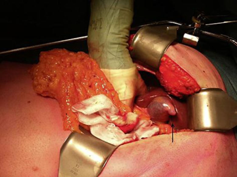
A Phrygian cap is a congenital anomaly of the gallbladder with an incidence of 4%. It can simulate a mass in the liver during hepatobiliary imaging and is sometimes mistaken for pathology. A Phrygian cap, however, has no pathological significance and normally causes no symptoms. A case will be presented where a Phrygian cap was found by coincidence during surgery. The patient was operated for colon cancer with liver metastasis in segment V. He underwent a simultaneous right hemicolectomy and wedge resection of the liver lesion. During perioperative inspection, a gallbladder with a folded fundus was seen. This deformity was, in retrospective, detected on the preoperative MRI scan. The patient underwent cholecystectomy to make the wedge resection easier to perform. Otherwise, cholecystectomy for a Phrygian cap is only indicated in case of symptoms. Radiographic imaging can be helpful in narrowing the differential diagnosis. To our knowledge, there is no recent literature about the Phrygian cap and its imaging aspects. Nowadays, multiphase MRI, or multiphase CT in case of MRI contraindication, are the first choices of hepatobiliary imaging.
Keywords: Cholecystectomy; Congenital anomaly; Gallbladder abnormalities; Gallbladder imaging.
Figures


References
-
- Hardy KJ. Carl Langenbuch and the Lazarus Hospital: events and circumstances surrounding the first cholecystectomy. Aust N Z J Surg. 1993;63:56–64. - PubMed
-
- Severn CB. A morphological study of the development of the human liver. 1. Development of the hepatic diverticulum. Am J Anat. 1971;131:133–158. - PubMed
-
- Rappaport AM, Wanless IR. Diseases of the Liver. ed 7. Philadelphia: Lippincott; 1993. p. 1.
-
- Lamah M, Karanjia ND, Dickson GH. Anatomical variations of the extrahepatic biliary tree: review of the world literature. Clin Anat. 2001;14:167–172. - PubMed
Publication types
LinkOut - more resources
Full Text Sources
Other Literature Sources
Miscellaneous
