

Beam hardening artifacts by dental implants: Comparison of cone-beam and 64-slice computed tomography scanners
- PMID: 24019808
- PMCID: PMC3760363
Beam hardening artifacts by dental implants: Comparison of cone-beam and 64-slice computed tomography scanners
Abstract
Background: Cone beam computed tomography (CBCT) is an alternative to a computed tomography (CT) scan, which is appropriate for a wide range of craniomaxillofacial indications. The long-term use of metallic materials in dentistry means that artifacts caused by metallic restorations in the oral cavity should be taken into account when utilizing CBCT and CT scanners. The aim of this study was to quantitatively compare the beam hardening artifacts produced by dental implants between CBCT and a 64-Slice CT scanner.
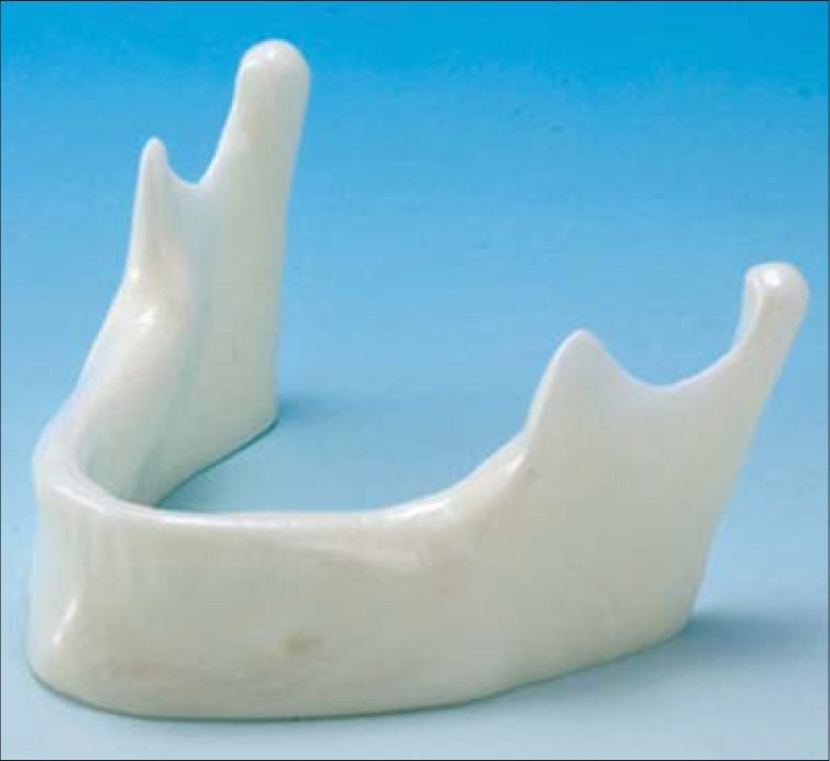
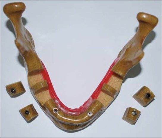
Materials and methods: In this descriptive study, an implant drilling model similar to the human mandible was used in the present study. The implants (Dentis) were placed in the canine, premolar and molar areas. Three series of scans were provided from the implant areas using Somatom Sensation 64-slice and NewTom VGi (CBCT) CT scanners. Identical images were evaluated by three radiologists. The artifacts in each image were determined based on pre-determined criteria. Kruskal-Wallis test was used to compare mean values; Mann-Whitney U test was used for two-by-two comparisons when there was a statistical significance (P < 0.05).
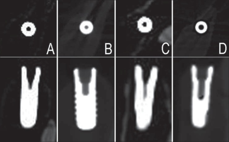
Results: The images of the two scanners had similar resolutions in axial sections (P = 0.299). In coronal sections, there were significant differences in the resolutions of the images produced by the two scanners (P < 0.001), with a higher resolution in the images produced by NewTom VGi scanner. On the whole, there were significant differences between the resolutions of the images produced by the two CT scanners (P < 0.001), with higher resolution in the images produced by NewTom VGi scanner in comparison to those of Somatom Sensation.
Conclusion: Given the high quality of the images produced by NewTom VGi and the lower costs in comparison to CT, the use of the images of this scanner in dental procedures is recommended, especially in patients with extensive restorations, multiple prostheses and previous implants.
Keywords: Beam hardening artifacts; cone beam computed tomography; dental implants.
Conflict of interest statement
Figures
References
-
- Chindasombatjareon J, Kakimoto N, Murakami S, Maeda Y, Furukawa S. Quantitative analysis of metalic artifacts caused by dental metals: Comparison of cone-beam and multi-detector row CT scanners. Oral Radiol. 2011;27:114–20.
-
- Ludlow JB, Davies-Ludlow LE, Brooks SL, Howerton WB. Dosimetry of 3 CBCT devices for oral and maxillofacial radiology: CB Mercuray, NewTom 3G and i-CAT. Dentomaxillofac Radiol. 2006;35:219–26. - PubMed
-
- Ludlow JB, Ivanovic M. Comparative dosimetry of dental CBCT devices and 64-slice CT for oral and maxillofacial radiology. Oral Surg Oral Med Oral Pathol Oral Radiol Endod. 2008;106:106–14. - PubMed
-
- Schulze RK, Berndt D, d’Hoedt B. On cone-beam computed tomography artifacts induced by titanium implants. Clin Oral Implants Res. 2010;21:100–7. - PubMed
-
- White SC, Pharoah MJ. Oral Radiology: Principles and Interpretation. 6th ed. St. Louis: Mosby; 2009. Cone- beam computed tomography; pp. 235–7.
LinkOut - more resources
Full Text Sources