

Case Reports
Oral malignant melanoma: A case report of an unusual clinical and histologic presentation
Affiliations
- PMID: 24019813
- PMCID: PMC3760368
Item in Clipboard
Case Reports
Oral malignant melanoma: A case report of an unusual clinical and histologic presentation
Dent Res J (Isfahan).
2013 May.
Abstract
Malignant melanoma is a potentially aggressive tumor of melanocytic origin. Primary oral malignant melanoma is a rare neoplasm, accounting for 0.5% of all oral malignancies. The present case occurred in a 60-year-old female patient, as a pedunculated growth involving the palate and alveolar ridge and histologically showing a desmoplastic differentiation. The article discusses the distinct clinico-pathologic presentation of this case and emphasizes on the need to identify and report such cases for further understanding of their biologic behavior.
Keywords: Desmoplastic melanoma; malignant melanoma; oral mucosal melanoma.
Conflict of interest statement
Figures

Clinical picture showing a pedunculated lesion involving the palate
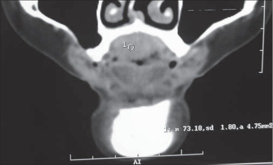
Computed tomography image showing subtle bony erosion noted over the mid portion of the hard palate

Superficial dermis showing pigmented epitheloid and spindle tumor cells (H and E, ×40)

Deep dermis showing the tumor with predominant spindle cell type arranged in irregular branching pattern with intervening fibrous septae (H and E, ×10)
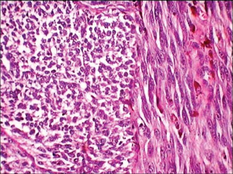
Spindle tumor cells appear fusiform to rounded in different orientations (H and E, ×40)

Tumor cells showing atypical features like mitosis, pleomorphism, hyperchromatism, increased nuclear cytoplasmic ratio and prominent nucleoli (H and E, ×40)
Similar articles
-
Desmoplastic malignant melanoma on the buttock of an 18-year-old girl: differentiation from desmoplastic nevus.Am J Dermatopathol. 1999 Apr;21(2):170-3. doi: 10.1097/00000372-199904000-00011. Am J Dermatopathol. 1999. PMID: 10218679
-
Primary malignant melanoma of oral cavity: A report of three rare cases.Contemp Clin Dent. 2016 Jan-Mar;7(1):87-9. doi: 10.4103/0976-237X.177094. Contemp Clin Dent. 2016. PMID: 27041909 Free PMC article.
-
A Large Oral Melanoma: A Case Report of a Rare but Aggressive Malignancy.Eur J Dent. 2021 Oct;15(4):812-816. doi: 10.1055/s-0041-1731836. Epub 2021 Aug 24. Eur J Dent. 2021. PMID: 34428837 Free PMC article.
-
Oral malignant melanoma--an unusual presentation.Gerodontology. 2012 Jun;29(2):e1241-3. doi: 10.1111/j.1741-2358.2010.00450.x. Epub 2010 Nov 17. Gerodontology. 2012. PMID: 22612841 Review.
-
Black and Brown Oro-facial Mucocutaneous Neoplasms.Head Neck Pathol. 2019 Mar;13(1):56-70. doi: 10.1007/s12105-019-01008-2. Epub 2019 Jan 29. Head Neck Pathol. 2019. PMID: 30693458 Free PMC article. Review.
Cited by
-
Primary amelanotic malignant melanoma of parotid and submandibular salivary gland: A rare case report.J Oral Maxillofac Pathol. 2022 Apr-Jun;26(2):263-267. doi: 10.4103/jomfp.jomfp_183_21. Epub 2022 Jun 28. J Oral Maxillofac Pathol. 2022. PMID: 35968175 Free PMC article.
-
Desmoplastic melanoma of the oral cavity: diagnostic pitfalls and clinical characteristics.J Korean Assoc Oral Maxillofac Surg. 2018 Apr;44(2):66-72. doi: 10.5125/jkaoms.2018.44.2.66. Epub 2018 Apr 25. J Korean Assoc Oral Maxillofac Surg. 2018. PMID: 29732311 Free PMC article.
References
-
- van der Waal RI, Snow GB, Karim AB, van der Waal I. Primary malignant melanoma of the oral cavity: A review of eight cases. Br Dent J. 1994;176:185–8. - PubMed
-
- Neville BD, Damm DD, Carl MA, Bouquot JE. Melanoma-Epithelial Pathology. 2nd ed. Philadelphia: Saunders; 2005. pp. 376–80.
-
- Gu GM, Epstein JB, Morton TH., Jr Intraoral melanoma: Long-term follow-up and implication for dental clinicians. A case report and literature review. Oral Surg Oral Med Oral Pathol Oral Radiol Endod. 2003;96:404–13. - PubMed
-
- Hicks MJ, Flaitz CM. Oral mucosal melanoma: Epidemiology and pathobiology. Oral Oncol. 2000;36:152–69. - PubMed
-
- Speight PM. Lyon: IARC Press; 2005. Mucosal malignant melanoma. World Health Organization Classification of Tumours, Pathology and Genetics of Head and Neck Tumours; pp. 206–7.
Publication types
LinkOut - more resources
Full Text Sources