

Altered expression of cyclin A 1 in muscle of patients with facioscapulohumeral muscle dystrophy (FSHD-1)
- PMID: 24019929
- PMCID: PMC3760810
- DOI: 10.1371/journal.pone.0073573
Altered expression of cyclin A 1 in muscle of patients with facioscapulohumeral muscle dystrophy (FSHD-1)
Abstract
Objectives: Cyclin A1 regulates cell cycle activity and proliferation in somatic and germ-line cells. Its expression increases in G1/S phase and reaches a maximum in G2 and M phases. Altered cyclin A1 expression might contribute to clinical symptoms in facioscapulohumeral muscular dystrophy (FSHD).
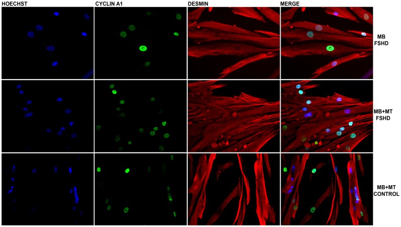
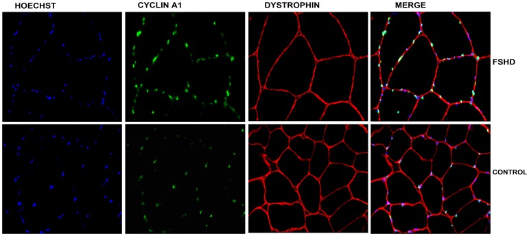
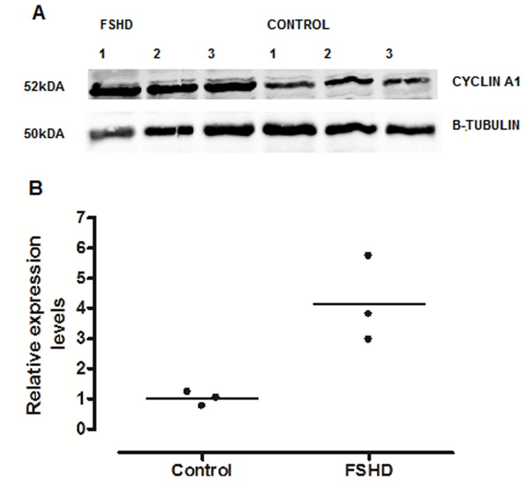
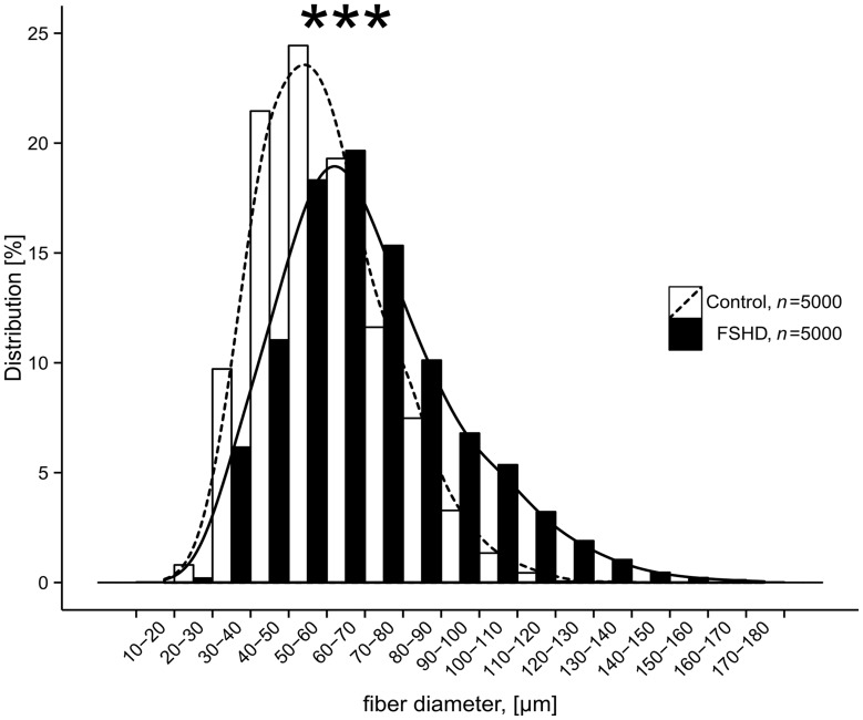
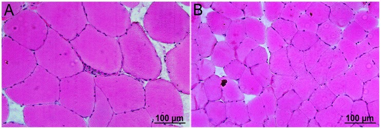
Methods: Muscle biopsies were taken from the Vastus lateralis muscle for cDNA microarray, RT-PCR, immunohistochemistry and Western blot analyses to assess RNA and protein expression of cyclin A1 in human muscle cell lines and muscle tissue. Muscle fibers diameter was calculated on cryosections to test for hypertrophy.
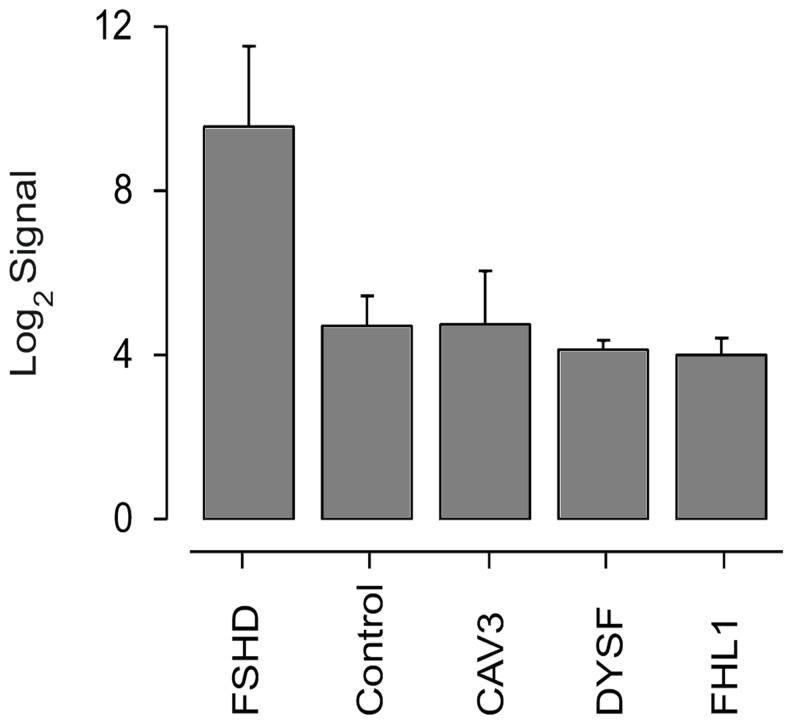
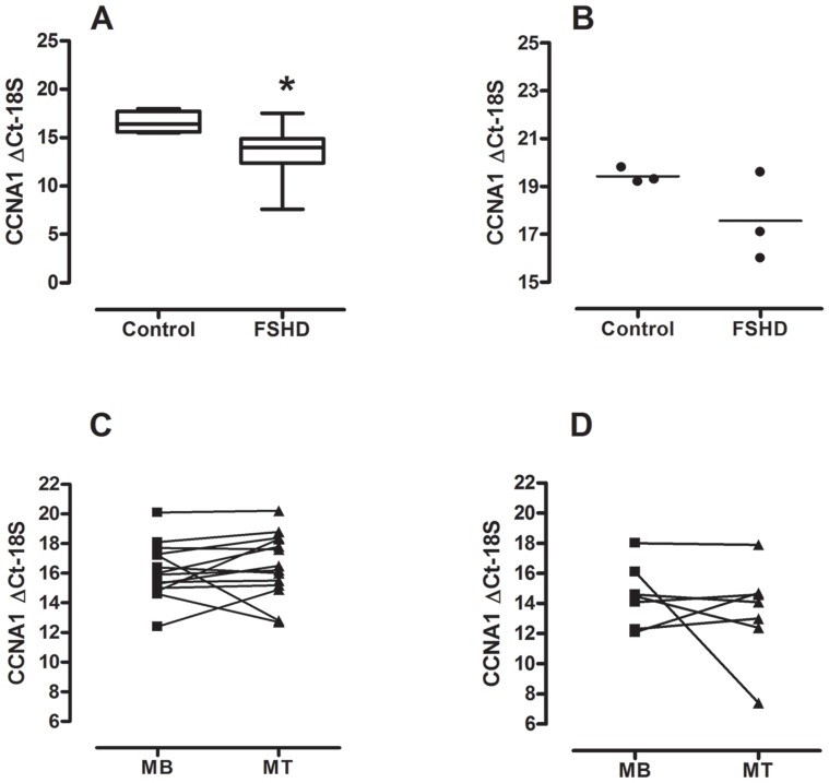
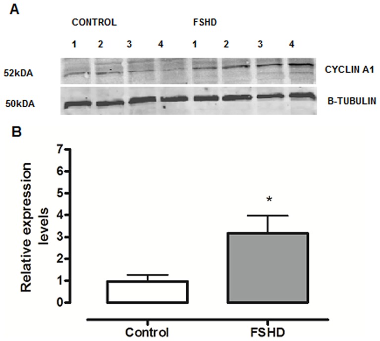
Results: cDNA microarray data showed specifically elevated cyclin A1 levels in FSHD vs. other muscular disorders such as caveolinopathy, dysferlinopathy, four and a half LIM domains protein 1 deficiency and healthy controls. Data could be confirmed with RT-PCR and Western blot analysis showing up-regulated cyclin A1 levels also at protein level. We found also clear signs of hypertrophy within the Vastus lateralis muscle in FSHD-1 patients.
Conclusions: In most somatic human cell lines, cyclin A1 levels are low. Overexpression of cyclin A1 in FSHD indicates cell cycle dysregulation in FSHD and might contribute to clinical symptoms of this disease.
Conflict of interest statement
Figures
References
-
- Gabellini D, D'Antona G, Moggio M, Prelle A, Zecca C, et al. (2006) Facioscapulohumeral muscular dystrophy in mice overexpressing FRG1. Nature 439: 973–977 nature04422 [pii];10.1038/nature04422 [doi] - DOI - PubMed
-
- Tawil R, Van Der Maarel SM (2006) Facioscapulohumeral muscular dystrophy. Muscle Nerve 34: 1–15 10.1002/mus.20522 [doi] - DOI - PubMed
-
- Tawil R (2008) Facioscapulohumeral muscular dystrophy. Neurotherapeutics 5: 601–606 S1933-7213(08)00139-6 [pii];10.1016/j.nurt.2008.07.005 [doi] - DOI - PMC - PubMed
-
- Tupler R, Gabellini D (2004) Molecular basis of facioscapulohumeral muscular dystrophy. Cell Mol Life Sci 61: 557–566 10.1007/s00018-003-3285-3 [doi] - DOI - PMC - PubMed
-
- Barat-Houari M, Nguyen K, Bernard R, Fernandez C, Vovan C, et al. (2010) New multiplex PCR-based protocol allowing indirect diagnosis of FSHD on single cells: can PGD be offered despite high risk of recombination? Eur J Hum Genet 18: 533–538 ejhg2009207 [pii];10.1038/ejhg.2009.207 [doi] - DOI - PMC - PubMed
Publication types
MeSH terms
Substances
LinkOut - more resources
Full Text Sources
Other Literature Sources
Molecular Biology Databases
Research Materials
