. 2013 Sep 3;8(9):e73636.
doi: 10.1371/journal.pone.0073636. eCollection 2013.

SGO1 maintains bovine meiotic and mitotic centromeric cohesions of sister chromatids and directly affects embryo development

Affiliations

SGO1 maintains bovine meiotic and mitotic centromeric cohesions of sister chromatids and directly affects embryo development

Feng-Xia Yin et al. PLoS One. .

Abstract

Shugoshin (SGO) is a critical factor that enforces cohesion from segregation of paired sister chromatids during mitosis and meiosis. It has been studied mainly in invertebrates. Knowledge of SGO(s) in a mammalian system has only been reported in the mouse and Hela cells. In this study, the functions of SGO1 in bovine oocytes during meiotic maturation, early embryonic development and somatic cell mitosis were investigated. The results showed that SGO1 was expressed from germinal vesicle (GV) to the metaphase II stage. SGO1 accumulated on condensed and scattered chromosomes from pre-metaphase I to metaphase II. The over-expression of SGO1 did not interfere with the process of homologous chromosome separation, although once separated they were unable to move to the opposing spindle poles. This often resulted in the formation of oocytes with 60 replicated chromosomes. Depletion of SGO1 in GV oocytes affected chromosomal separation resulting in abnormal chromosome alignment at a significantly higher proportion than in control oocytes. Knockdown of SGO1 expression significantly decreased the embryonic developmental rate and quality. To further confirm the function(s) of SGO1 during mitosis, bovine embryonic fibroblast cells were transfected with SGO1 siRNAs. SGO1 depletion induced the premature dissociation of chromosomal cohesion at the centromere and along the chromosome arm giving rise to abnormal appearing mitotic patterns. The results of this study infer that SGO1 is involved in the centromeric cohesion of sister chromatids and chromosomal movement towards the spindle poles. Depletion of SGO1 causes arrestment of cell division in meiosis and mitosis.


Conflict of interest statement

Competing Interests: The authors have declared that no competing interests exist.

Figures

Figure 1
Figure 1. The relative mRNA levels of SGO1 in a matured bovine oocyte from GV to MII stages.
The expression level of SGO1 at GV stage (0 h) was adjusted to 100%. Different superscripts indicate a statistical difference (P<0.01).
Figure 2
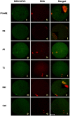
Figure 2. The localization of SGO1-MYC during bovine oocyte maturation.
Oocytes matured at different stages were stained with c-myc antibody and PI. Green, SGO1-MYC; red, DNA; yellow, overlapping of red and green. pre-MI, pre-metaphase I; MI, metaphase I; AI, anaphase I; TI, telophase I; MII, metaphase II. Bar = 50 µm; original magnification×400.
Figure 3
Figure 3. Over-expression of exogenously delivered SGO1 in bovine oocytes during meiosis inhibited chromosome polarization.
A) Images of oocytes without PB1 after SGO1 over-expression. Oocytes were stained with c-myc antibody and PI. Green, SGO1-MYC; red, DNA; yellow, overlapping of red and green. Bar = 20 µm; original magnification×400.B) Representative chromosome configurations are shown. I, metaphase I; II, 60 chromosomes; III, anaphase I; IV, metaphase II. Bar = 5 µm; original magnification×1000. C) Corresponding figure presentation as in B. The error bar is expressed as mean ± SEM.* indicate a statistical difference (P<0.01).
Figure 4
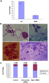
Figure 4. Depletion of bovine SGO1 in GV oocytes affected chromosome separation.
A) SGO1 mRNA level after RNAi. Different superscripts indicate a statistical difference (P<0.01). The error bar is expressed as mean ± SEM. B) Chromosome spread after SGO1 RNAi. I: normal metaphase II chromosomes; II, multi-polar chromosome; III and IV, abnormal chromosomal separation, bar = 10 µm; original magnification×1000. C) The percentage of abnormal chromosomes (III and IV) after SGO1-specific interference. Different superscripts indicate a statistical difference (P<0.05). The error bar is expressed as mean ± SEM.
Figure 5
Figure 5. Assessment of cell number of the resulted bovine blastocysts derived from SGO1-RNAi embryos.
The arrows show fragmented nuclei. Stained with Hoechst 33342. Bar = 50 µm; original magnification×400.
Figure 6
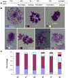
Figure 6. SGO1 depletion in bovine mitosis caused various types of mitotic arrestment.
A) Schematic overview of experimental process. B) Representative images of seven kinds of mitotic patterns. I: prophase. II: metaphase and metaphase-like. III: anaphase. IV: Early separation of sister chromatid. Some or all arm cohesion of the chromosomes seems to be lost and displays a “V” appearance. V: At this stage, centromeric cohesion appears nonexistent. Some or all sister chromatids are still aligned in pairs. VI: Chromosomal composition appears as in V, but is hyper condensed state. VII: Sister chromatids are completely separated and hyper condensed, individual chromatids are scattered disorderly. Bar = 10 µm; original magnification×1000. C) Percentages of each kind of mitotic cells showed. c5, c7, c9 stand for non-sense siRNA treated cells harvest at 5 h, 7 h and 9 h after release 2, t5, t7, t9 stand for SGO1 specific siRNA treated cells harvest at 5 h, 7 h and 9 h after siRNA treatment.

Similar articles

Cited by

References

    1. Petronczki M, Siomos MF, Nasmyth K (2003) Un menage a quatre: the molecular biology of chromosome segregation in meiosis. Cell 112: 423–440. - PubMed
    1. Watanabe Y (2005) Shugoshin: guardian spirit at the centromere. Curr Opin Cell Biol 17: 590–595. - PubMed
    1. Gutié rrez-Caballero C, Cebollero LR, Pendá s AM (2012) Shugoshins: from protectors of cohesion to versatile adaptors at the centromere. Trends Genet 28(7): 351–360. - PubMed
    1. Pouwels J, Kukkonen AM, Lan WJ, Daum JR, Gorbsky GJ, et al. (2007) Shugoshin 1 plays a central role in kinetochore assembly and is required for kinetochore targeting of Plk1. Cell Cycle 6: 1579–1585. - PubMed
    1. Tang Z, Sun Y, Harley SE, Zou H, Yu H (2004) Human Bub1 protects centromeric sister-chromatid cohesion through Shugoshin during mitosis. PNAS 101: 18012–18017. - PMC - PubMed

Publication types

LinkOut - more resources