. 2013 Sep 3;8(9):e73701.
doi: 10.1371/journal.pone.0073701. eCollection 2013.

Beetroot-pigment-derived colorimetric sensor for detection of calcium dipicolinate in bacterial spores

Affiliations

Beetroot-pigment-derived colorimetric sensor for detection of calcium dipicolinate in bacterial spores

Letícia Christina Pires Gonçalves et al. PLoS One. .

Abstract

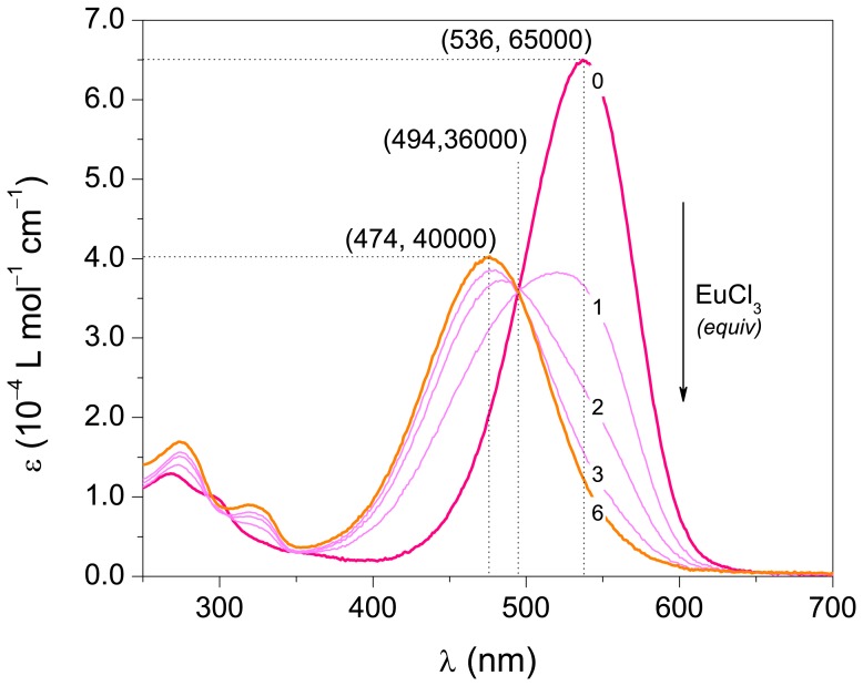
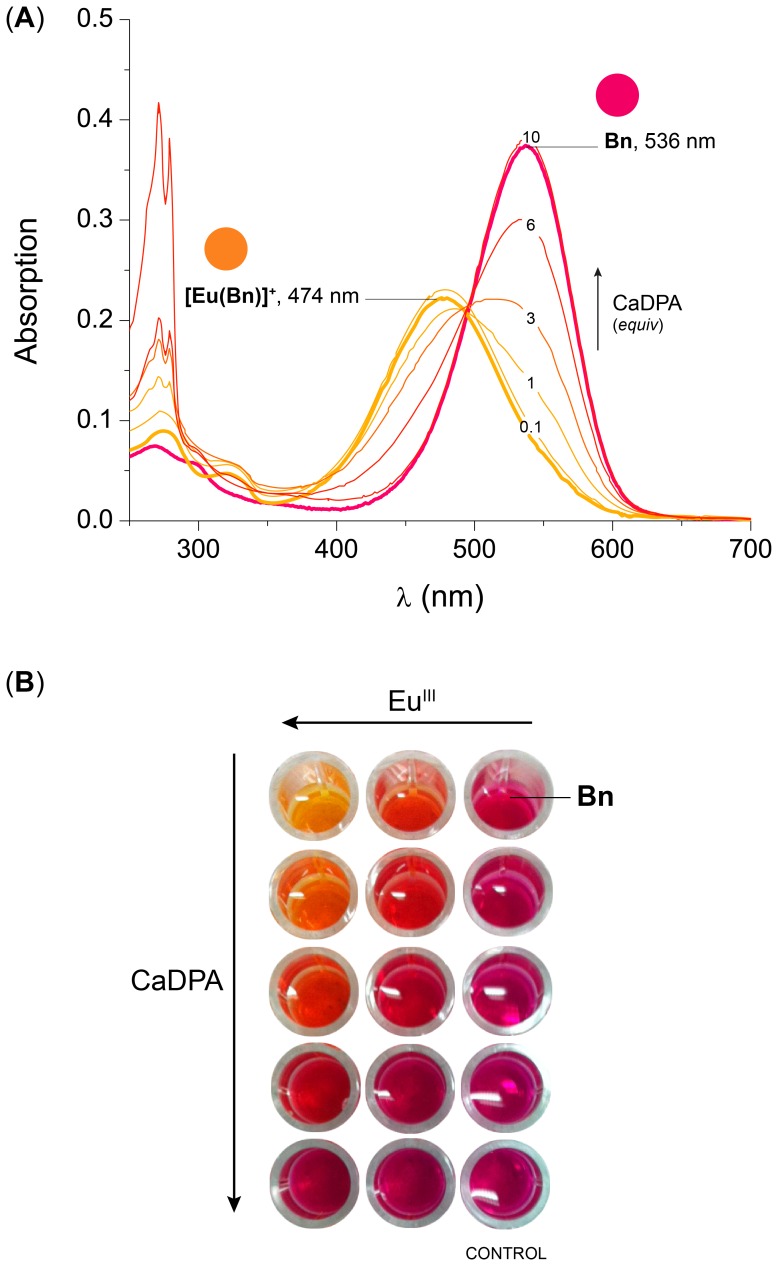
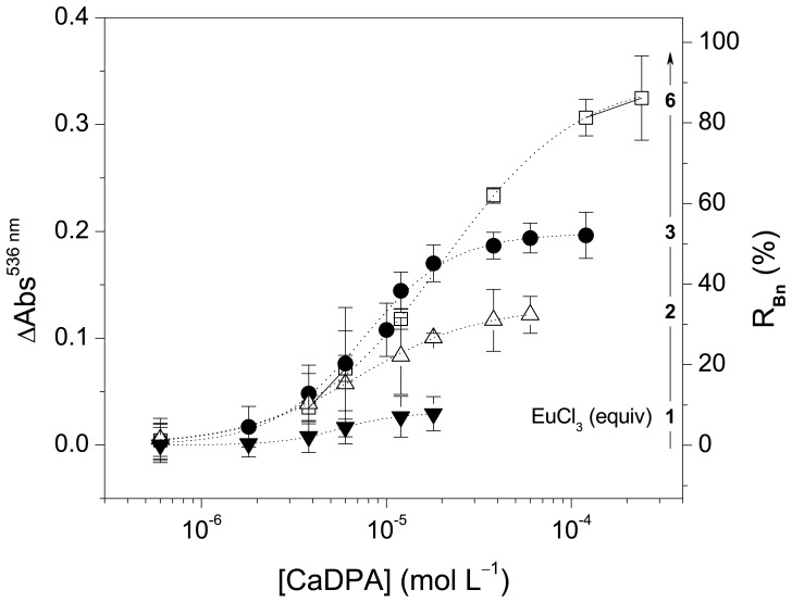
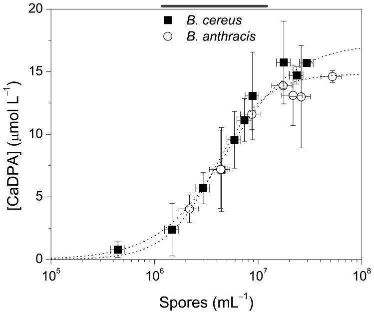
In this proof-of-concept study, we describe the use of the main red beet pigment betanin for the quantification of calcium dipicolinate in bacterial spores, including Bacillus anthracis. In the presence of europium(III) ions, betanin is converted to a water-soluble, non-luminescent orange 1∶1 complex with a stability constant of 1.4 × 10(5) L mol(-1). The addition of calcium dipicolinate, largely found in bacterial spores, changes the color of the aqueous solution of [Eu(Bn)(+)] from orange to magenta. The limit of detection (LOD) of calcium dipicolinate is around 2.0 × 10(-6) mol L(-1) and the LOD determined for both spores, B. cereus and B. anthracis, is (1.1 ± 0.3)× 10(6) spores mL(-1). This simple, green, fast and low cost colorimetric assay was selective for calcium dipicolinate when compared to several analogous compounds. The importance of this work relies on the potential use of betalains, raw natural pigments, as colorimetric sensors for biological applications.


Conflict of interest statement

Competing Interests: The authors have declared that no competing interests exist.

Figures

Figure 1
Figure 1. Absorptivity profile for the formation of [Eu(Bn)]+ by the addition of EuCl3 to a solution of Bn in MOPS buffer pH = 7.5.
The numbers in the curves indicate the concentration of EuCl3 in equivalents of Bn.
Figure 2
Figure 2. Effect of the addition of CaDPA on an aqueous solution of [Eu(Bn)]+.
(A) Absorption profile for the formation of Bn by the addition of CaDPA to a solution of [Eu(Bn)]+; The numbers in the curves indicate the concentration of CaDPA in equivalents of Bn. (B) Picture of a microplate containing Bn and increasing amounts of EuCl3 and CaDPA, the background was removed for clarity. [Bn] = 5.8 µmol L–1, [EuCl3] = 34.8 µmol L–1 (6 equiv) in MOPS buffer pH = 7.5.
Figure 3
Figure 3. Effect of the concentration of EuIII on the quantification of CaDPA.
The variation of the absorbance at 536 nm and the percent recovery of Bn are plotted against the concentration of CaDPA (log scale). Lines indicate the fitting of data using Eq. (3) with parameter Amin set to zero; [Bn] = 5.8 µmol L–1 in MOPS buffer. LOD are: 6 equiv, (2.8±1.5)×10–6 mol L–1; 3 equiv, (2.2±1.1)×10–6 mol L–1; 2 equiv, (1.4±0.9)×10–6 mol L–1 and 1 equiv, (1.7±0.8)×10–6 mol L–1.
Figure 4
Figure 4. CaDPA concentration (log scale) plotted against the variation in the absorbance at 536 nm (ΔAbs536 nm) and the normalized response (N Bn).
The diagonal straight-line shows when the plotted parameters are linearly correlated. Curved lines are the confidence bands at the 95% level. Error bars represent the sd of triplicates. [Bn] = 5.8 µmol L–1, [EuCl3] = 17.4 µmol L–1 (3 equiv).
Figure 5
Figure 5. Quantification of bacteria of the genus Bacillus by the amount of CaDPA released upon thermal treatment.
Vertical and horizontal error bars indicate uncertainties in spore counting and CaDPA quantification (sd, N = 3). The concentration of spores is in the log scale for clarity. [Bn] = 5.8 µmol L–1, [EuCl3] = 17.4 µmol L–1 (3 equiv) in MOPS buffer pH = 7.5.

Similar articles

Cited by

References

    1. Vilas-Boas GT, Peruca APS, Arantes OMN (2007) Biology and taxonomy of Bacillus cereus, Bacillus anthracis, and Bacillus thuringiensis. Can J Microbiol 53: 673–687. - PubMed
    1. Inglesby TV, Henderson DA, Bartlett JG, Ascher MS, Eitzen E, et al. (1999) Anthrax as a biological weapon: Medical and public health management. JAMA, J Am Med Assoc 281: 1735–1745. - PubMed
    1. Inglesby TV, O’Toole T, Henderson DA, Bartlett JG, Ascher MS, et al. (2002) Anthrax as a biological weapon, 2002: Updated recommendations for management. JAMA, J Am Med Assoc 287: 2236–2252. - PubMed
    1. Sanderson WT, Stoddard RR, Echt AS, Piacitelli CA, Kim D, et al. (2004) Bacillus anthracis contamination and inhalational anthrax in a mail processing and distribution center. J Appl Microbiol 96: 1048–1056. - PubMed
    1. Dragon DC, Rennie RP (1995) The ecology of anthrax spores: tough but not invincible. Can Vet J 36: 295–301. - PMC - PubMed

Publication types

MeSH terms

LinkOut - more resources