. 2013 Sep 3;8(9):e74328.
doi: 10.1371/journal.pone.0074328. eCollection 2013.

Experimental pathways towards developing a rotavirus reverse genetics system: synthetic full length rotavirus ssRNAs are neither infectious nor translated in permissive cells

Affiliations

Experimental pathways towards developing a rotavirus reverse genetics system: synthetic full length rotavirus ssRNAs are neither infectious nor translated in permissive cells

James E Richards et al. PLoS One. .

Abstract

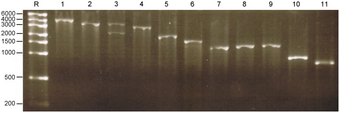
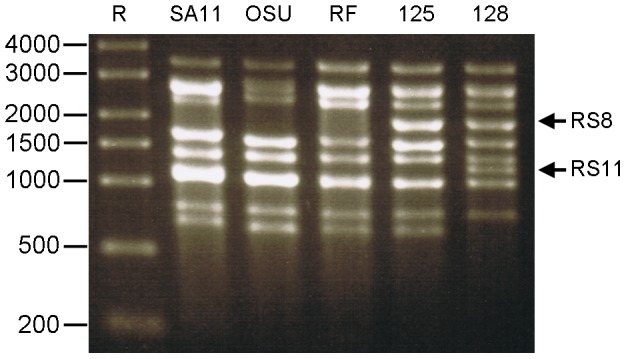
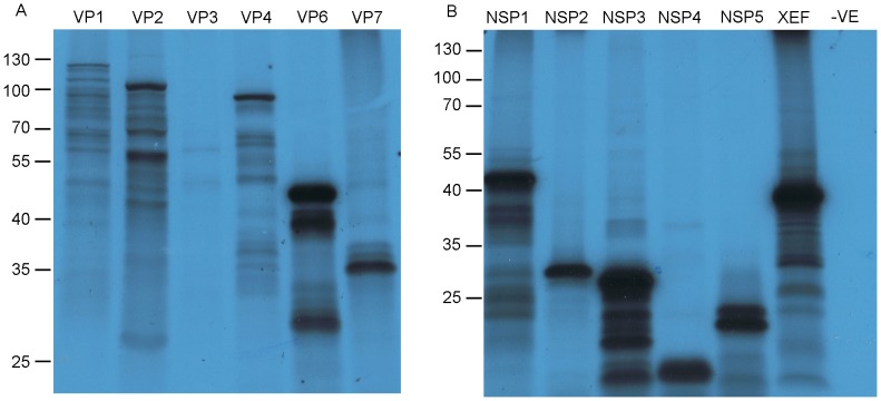
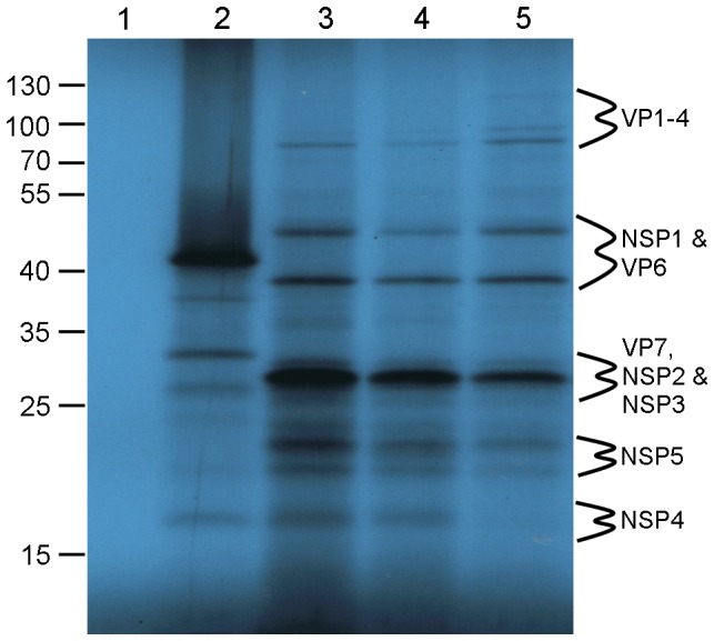
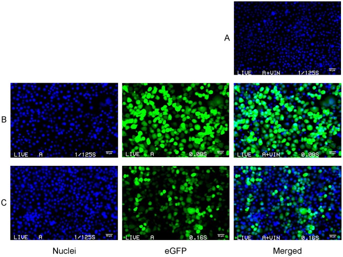
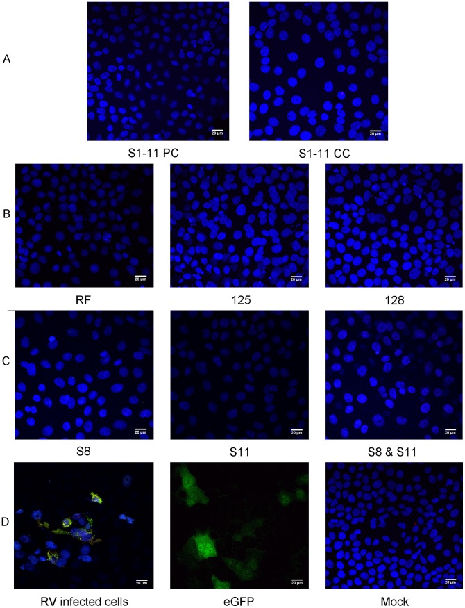
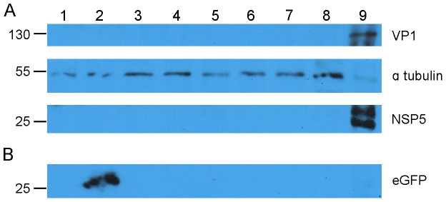
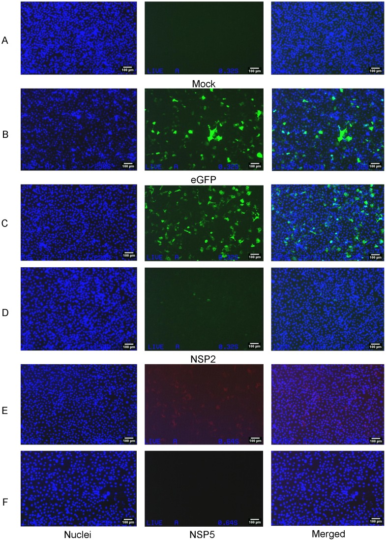
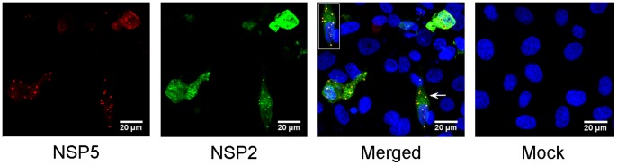
At present the ability to create rationally engineered mutant rotaviruses is limited because of the lack of a tractable helper virus-free reverse genetics system. Using the cell culture adapted bovine RV RF strain (G6P6 [1]), we have attempted to recover infectious RV by co-transfecting in vitro transcribed ssRNAs which are identical in sequence to the positive sense strand of each of the 11 dsRNA genomic segments of the RF strain. The RNAs were produced either from cDNAs cloned by a target sequence-independent procedure, or from purified double layered RV particles (DLPs). We have validated their translational function by in vitro synthesis of (35)S-labelled proteins in rabbit reticulocyte lysates; all 11 proteins encoded by the RV genome were expressed. Transfection experiments with DLP- or cDNA-derived ssRNAs suggested that the RNAs do not act independently as mRNAs for protein synthesis, once delivered into various mammalian cell lines, and exhibit cytotoxicity. Transfected RNAs were not infectious since a viral cytopathic effect was not observed after infection of MA104 cells with lysates from transfected cells. By contrast, an engineered mRNA encoding eGFP was expressed when transfected under identical conditions into the same cell lines. Co-expression of plasmids encoding NSP2 and NSP5 using a fowlpox T7 polymerase recombinant virus revealed viroplasm-like structure formation, but this did not enable the translation of transfected RV ssRNAs. Attempts to recover RV from ssRNAs transcribed intracellularly from transfected cDNAs were also unsuccessful and suggested that these RNAs were also not translated, in contrast to successful translation from a transfected cDNA encoding an eGFP mRNA.


Conflict of interest statement

Competing Interests: The authors have declared that no competing interests exist.

Figures

Figure 1
Figure 1. Agarose gel electrophoresis of the in vitro transcribed ssRNAs of the 11 RF RV segments.
In vitro transcribed viral positive sense RNA, transcribed in the presence of a cap analogue from linear templates with a RE digested 3′ end. 200 ng of each ssRNA transcript was loaded onto the gel; 1.5% TBE AGE 80 V for 45 min. Lane R: RiboRuler™ High Range RNA markers (in bases); lanes 1–11: positive sense co-capped RV RF ssRNAs corresponding to segments 1 to 11, respectively.
Figure 2
Figure 2. ssRNA derived from several strains of RV DLPs.
ssRNA were synthesized in vitro from purified RV DLPs. DLPs were incubated at 37°C for 2 hours with the necessary components for positive sense ssRNA synthesis. 1.5% TBE AGE 45 V for 120 min. Lane R: RiboRuler™ High Range (in bases); 1.5 µg ssRNA derived from DLPs of RV strains SA11, OSU, RF, 125 and 128, respectively. DLP-derived ssRNAs 2 and 3 comigrate in all samples., ssRNAS 7–9 comigrate in SA11, OSU and RF, but only ssRNAS 7 and 9 comigrate in 125 and 128. Arrows indicate the altered migration of ssRNAs synthesised from the templates of rearranged genomic segments (RS). RS8, RS11 rearranged segment 8 or 11, respectively, of RV strains 125 and 128.
Figure 3
Figure 3. eGFP ssRNA species produced in vitro.
ssRNAs were synthesised from PCR-derived amplicons with a T7 Pol promoter introduced at the 5′ end to facilitate in vitro transcription. PCR amplicons were digested with BsmBI to define the 3′ end of the transcription cassette. Templates, either 500 ng or 1 µg, were incubated with T7 Pol in the absence or presence of a cap analogue using MEGAscript® or Mmessage Mmachine®. Uncapped ssRNAs were purified and post-transcriptionally capped using ScriptCap™ m7G Capping System. ssRNA was polyadenylated using E. coli polyadenylation polymerase (ePAP) Ambion. Lane R: RiboRuler™ High Range; lanes 1–6: approximately 250 ng each of eGFP ssRNA; uncapped, post-capped, uncapped polyadenylated, post-capped polyadenylated, co-capped, co-capped polyadenylated, respectively. 1.5% TBE AGE 60 V for –90 min. The polyadenylated RNA bands are less sharp as the molecules differ in the numbers of A residues added at the 3′end.
Figure 4
Figure 4. In vitro translation of RV proteins using S1 - 11 in vitro transcribed ssRNAs.
In vitro translation using all in vitro transcribed RV ssRNAs as templates for protein synthesis. 500 ng of ssRNA was incubated in RRL with 35S L-methionine for 3 hours and analysed using SDS-PAGE. The dried gel was exposed to X-ray film for 36 hours. Panel A, 15% SDS - PAGE, contains the in vitro translated RV structural proteins; VP1, VP2, VP3, VP4, VP6 and VP7, respectively. Panel B, 12% SDS-PAGE, contains the in vitro translated RV non-structural proteins; NSP1, NSP2, NSP3, NSP4 and NSP5, respectively. Lane 6: Xenopus elongationfactor 1α (XEF) (positive control); lane 7: no ssRNA, (negative control). The sizes of protein markers run alongside during SDS-PAGE are indicated in kDa to the left of the autoradiographs.
Figure 5
Figure 5. In vitro translation of RV proteins from cohorts of cDNA-derived and DLP-derived ssRNAs.
In vitro translation of RV DLP derived ssRNAs. 1 µg of capped DLP ssRNA was incubated in a RRL as described, electrophoresed on a 15% SDS-PAGE alongside PageRuler™ protein markers (in kDa) and exposed to X-ray film for 4 days. Lane 1: no ssRNA; lane 2: XEF ssRNA; lanes 3 & 4: S1–11 post-capped or co-capped ssRNA respectively; lane 5: RV ssRNAs from RV RF strain DLPs. The sizes of protein markers run alongside SDS-PAGE are indicated in kDa to the right hand side and were used to predict the location of RV proteins.
Figure 6
Figure 6. Comparison of fluorescence between transfected eGFP RNA species.
COS-7 cells at 80% confluence, incubated in Opti-MEM I media were transfected with eGFP ssRNA using Lipofectamine 2000. Cells were stained with Hoechst 33342 and live imaged in an epifluorescence microscope at 24 hours post transfection. Panel A: mock transfected cells. Panels B & C: cells were transfected with 750 ng of eGFP ssRNA either post-capped or co-capped, respectively. Cells were exposed to UV (nuclei staining), blue light (eGFP excitation) or merged, respectively.
Figure 7
Figure 7. Transfection of COS-7 cells with in vitro transcribed ssRNAs encoding RV ssRNAs and DLP derived ssRNAs.
COS-7 cells at 80% confluence were transfected with ssRNAs encoding RV proteins using Mirus transfection reagent. Cells were fixed at 24 hours post transfection and stained with NSP2 and NSP5-specific antibodies (Table S4). Images were analysed by confocal microscopy. Panel A: transfection with cohorts of in vitro transcribed ssRNAs, 1 µg of S1– S11, post-capped (PC) or co-capped (CC) respectively, stained for both NSP2 and NSP5. Panel B: 1 µg of DLP derived ssRNAs from RV strains RF, 125 and 128 respectively, stained for both NSP2 and NSP5. Panel C: individual or co-transfection of 500 ng of ssRNAs S8, S11 or both stained for NSP2, NSP5 or both, respectively. Panel D: immunofluorescence of control MA104 cells infected with RF RV, stained for NSP2 and NSP5, transfection control with 1 µg of eGFP ssRNAs (autofluorescence), mock transfection stained for NSP2 and NSP5. Cell nuclei were stained with Hoechst 33342 in all panels. Scale bars: 20 µm.
Figure 8
Figure 8. Absence of VP1 and NSP5 expression from transfected ssRNAs by Western blotting.
COS-7 cells were transfected with 1 µg of RV ssRNA using the Mirus TransIT™ mRNA transfection reagent. Expression of RV proteins from cell lysates was sought by Western blot. Panel A, the membrane was split into three sections, to ascertain the presence of VP1, 170–70 kDa, loading control α tubulin, 70 - 40 kDa and NSP5, 40–15 kDa. Each section was incubated with the respective primary and secondary horseradish peroxidase (HRP) conjugated antibody (Table S5). Panel B, corresponding to the 40–15 KDa portion of membrane A which was reprobed for eGFP (Table S5). Proteins were visualised using the ECL Western blot detection reagents, light sensitive film was exposed to membranes for varying lengths of time depending on band intensity. Lane 1: mock; lanes 2–8 in vitro transcribed or DLP derived ssRNAs of: 2: eGFP, 3: S1– s11, 4: S1, 5: S11, 6: DLP RF, 7: S1 polyadenylated, 8: S11 polyadenylated; 9: COS-7 infected RV RF cell lysate.
Figure 9
Figure 9. FPV-T7-driven intracellular transcription and translation from transfected linear DNA templates of NSP2 and NSP5.
COS-7 cells infected with FPV-T7 Pol for 1 hour and transfected with 1 µg of linear PCR amplicon encoding either RV segments 8 or 11 or EGFP. pcDNA3-NSP2 and pT7V-NSP5 were used as protein controls. Cells were incubated for 24 hours, prior to fixing and staining for RV proteins as previously described. Cells transfected with EGFP were only stained with Hoechst 33342. Samples were imaged using a fluorescence microscope. All panels were exposed to UV light, panels A, B, C, and D were exposed to blue and panels E and F were exposed to green wavelengths of light. Panel A: mock transfection; panel B: transfection with T7EGFP; panel C: transfection with pcDNA3-NSP2; panel D: transfection with T7RFS8; panel E: transfection with pT7V-NSP5; panel F: transfection with T7RFS11. Cell nuclei were stained with Hoechst 33342. Scale bars: 100 µm.
Figure 10
Figure 10. Viroplasm-like structure (VLS) formation.
MA104 cells were infected with FPV-T7 for one hour before co-transfection with pT7V-NSP2 and pT7V-NSP5 plasmids using Lipofectamine 2000. Cells were fixed at 24 hours post transfection and stained for NSP2 and NSP5. Cell nuclei were stained with Hoechst 33342. VLS and mock panels are merged images of UV and the other two composite excitations for NSP2 or NSP5. Images were visualised by confocal microscopy. Arrows indicate VLS and location of enlarged inset images. Scale bars: 20 µm.

Similar articles

Cited by

References

    1. Estes MK, Kapikian AZ (2007) Rotaviruses. In: Knipe DM, Howley PM, editors. Fields Virology. 5th ed: Lippincott Williams & Wilkins/Wolters Kluwer, Philadelphia. 1917–1974.
    1. Tate JE, Burton AH, Boschi-Pinto C, Steele AD, Duque J, et al. (2012) 2008 estimate of worldwide rotavirus-associated mortality in children younger than 5 years before the introduction of universal rotavirus vaccination programmes: a systematic review and meta-analysis. The Lancet Infectious Diseases 12: 136–141. - PubMed
    1. Vesikari T, Matson DO, Dennehy P, Van Damme P, Santosham M, et al. (2006) Safety and Efficacy of a Pentavalent Human-Bovine (WC3) Reassortant Rotavirus Vaccine. New England Journal of Medicine 354: 23–33. - PubMed
    1. Ruiz-Palacios GM, Perez-Schael I, Velazquez FR, Abate H, Breuer T, et al. (2006) Safety and efficacy of an attenuated vaccine against severe rotavirus gastroenteritis. New England Journal of Medicine 354: 11–22. - PubMed
    1. SAGE (2009) Meeting of the immunization strategic advisory group of experts, April 2009 - conclusions and recommendations. Weekly Epidemiological Record 84: 220–235. - PubMed

Publication types