

doi: 10.4056/sigs.3276687.
eCollection 2013.
Non contiguous-finished genome sequence and description of Peptoniphilus obesi sp. nov
Affiliations
- PMID: 24019985
- PMCID: PMC3764929
- DOI: 10.4056/sigs.3276687
Item in Clipboard
Non contiguous-finished genome sequence and description of Peptoniphilus obesi sp. nov
Stand Genomic Sci.
.
Abstract
Peptoniphilus obesi strain ph1(T) sp. nov., is the type strain of P. obesi sp. nov., a new species within the genus Peptoniphilus. This strain, whose genome is described here, was isolated from the fecal flora of a 26-year-old woman suffering from morbid obesity. P. obesi strain ph1(T) is a Gram-positive, obligate anaerobic coccus. Here we describe the features of this organism, together with the complete genome sequence and annotation. The 1,774,150 bp long genome (1 chromosome but no plasmid) contains 1,689 protein-coding and 29 RNA genes, including 5 rRNA genes.
Keywords: Peptoniphilus obesi; genome.
Figures

Phylogenetic tree highlighting the position of Peptoniphilus obesi strain ph1T relative to a selection of type strains of validly published species of Peptoniphilus . GenBank accession numbers are indicated in parentheses. Sequences were aligned using CLUSTALW, and phylogenetic inferences obtained using the maximum-likelihood method within the MEGA software. Numbers at the nodes are percentages of bootstrap values obtained by repeating the analysis 500 times to generate a majority consensus tree. Peptoniphilus timonensis sp. nov., a new species that we recently proposed, was also included in the analysis [12]. Anaerococcus prevotii was used as outgroup. The scale bar represents a 2% nucleotide sequence divergence.

Gram staining of P. obesi strain ph1T
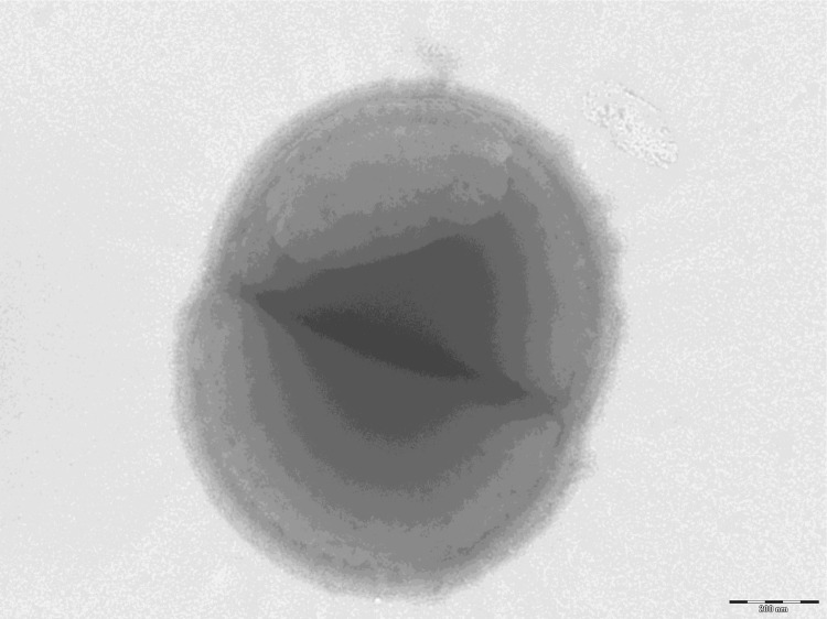
Transmission electron microscopy of P. obesi strain ph1T, using a Morgani 268D (Philips) at an operating voltage of 60kV. The scale bar represents 200 nm.

Reference mass spectrum from P. obesi strain ph1T. Spectra from 12 individual colonies were compared and a reference spectrum was generated.

Gel view comparing Peptoniphilus obesi ph1T spectra with other members into Peptoniphilus genera (Peptoniphilus timonensis , Peptoniphilus senegalensis, Peptoniphilus grossensis, Peptoniphilus ivorii , Peptoniphilus indolicus , Peptoniphilus harei , Peptoniphilus gorbachii and Peptoniphilus asaccharolyticus ). The Gel View displays the raw spectra of all loaded spectrum files arranged in a pseudo-gel like look. The x-axis records the m/z value. The left y-axis displays the running spectrum number originating from subsequent spectra loading. The peak intensity is expressed by a Gray scale scheme code. The color bar and the right y-axis indicate the relation between the color a peak is displayed with and the peak intensity in arbitrary units.

Graphical circular map of the chromosome. From the outside in, the outer two circles show open reading frames oriented in the forward (colored by COG categories) and reverse (colored by COG categories) directions, respectively. The third circle marks the rRNA gene operon (red) and tRNA genes (green). The fourth circle shows the G+C% content plot. The inner-most circle shows GC skew, purple indicating negative values whereas olive for positive values.
References
-
- Lagier JC, Armougom F, Million M, Hugon P, Pagnier I, Robert C, Bittar F, Fournous G, Gimenez G, Maraninchi M, et al. Microbial culturomics: paradigm shift in the human gut microbiome study. Clin Microbiol Infect 2012; 18:1185-1193 - PubMed
-
- Stackebrandt E, Ebers J. Taxonomic parameters revisited: tarnished gold standards. Microbiol Today 2006; 33:152-155
-
- Genome Online Database http://www.genomesonline.org/cgi-bin/GOLD/index.cgi
LinkOut - more resources
Full Text Sources
Molecular Biology Databases
