. 2014 May:50:12-22.
doi: 10.1016/j.jaut.2013.08.003. Epub 2013 Sep 7.

IL-27p28 inhibits central nervous system autoimmunity by concurrently antagonizing Th1 and Th17 responses

Affiliations

IL-27p28 inhibits central nervous system autoimmunity by concurrently antagonizing Th1 and Th17 responses

Wai Po Chong et al. J Autoimmun. 2014 May.

Abstract

Central nervous system (CNS) autoimmunity such as uveitis and multiple sclerosis is accompanied by Th1 and Th17 responses. In their corresponding animal models, experimental autoimmune uveitis (EAU) and experimental autoimmune encephalomyelitis (EAE), both responses are induced and can drive disease independently. Because immune responses have inherent plasticity, therapeutic targeting of only one pathway could promote the other, without reducing pathology. IL-27p28 antagonizes gp130, required for signaling by IL-27 and IL-6, which respectively promote Th1 and Th17 responses. We therefore examined its ability to protect the CNS by concurrently targeting both effector responses. Overexpression of IL-27p28 in vivo ameliorated EAU as well as EAE pathology and reduced tissue infiltration by Th1 and Th17 cells in a disease prevention, as well as in a disease reversal protocol. Mechanistic studies revealed inhibition of Th1 and Th17 commitment in vitro and decreased lineage stability of pre-formed effectors in vivo, with reduction in expression of gp130-dependent transcription factors and cytokines. Importantly, IL-27p28 inhibited polarization of human T cells to the Th1 and Th17 effector pathways. The ability of IL-27p28 to inhibit generation as well as function of pathogenic Th1 and Th17 effector cells has therapeutic implications for controlling immunologically complex autoimmune diseases.

Keywords: Autoimmunity; IL-27p28; gp130.


Figures

Fig. 1
Fig. 1
Overexpression of IL-27p28 suppresses EAU and EAE and reduces Th1 and Th17 immune responses. (A–B) p28-TG mice and WT littermates were immunized for EAU. (A) Average disease score and representative histopathology (day 21, hematoxylin-eosin, original magnification ×200). (B) Expression of IFN-γ, IL-17A and GM-CSF in CD4+ T cells from the eyes by intracellular staining on day 21. (C–D) B10.RIII mice were immunized for EAU and treated hydrodynamically with 20 μg of pmIL27p28 (d 0 and 7). (C) Average disease scores and representative histopathology (day 21, h&e, ×200). (D) Expression of IFN-γ, IL-17A and GM-CSF by CD4+ cells in uveitic eyes by intracellular staining (d 21). (E) C57BL/6 mice were immunized for EAU and treated with IL-27p28 (5 μg every other day) or PBS, starting from day 0 (9-10 mice per group), data shown is the average disease scores on day 21. (F) Anti-IRBP antibody titers in p28-TG mice and their WT littermates immunized for EAU. (G) EAE scores in IL-27p28 Tg mice and WT littermates immunized with MOG35–55. (H) EAE in WT C57BL/6 mice immunized with MOG35–55 and hydrodynamically injected with pmIL27p28 (d 0 and 7). (A, C, G, H) Data shown as mean ± SEM from 2 independent experiments with total of at least 10 mice per group. *: p < 0.05.
Fig. 2
Fig. 2
IL-27p28 suppresses IL-27, but not IL-12, driven Th1 responses. (A) CD4+CD62L+ cells from p28-TG mice or from WT littermates (with or without added IL-27p28) were stimulated with anti-CD3/CD28 in the presence of IL-12 or IL-27 for 3 days. (B) The same cells were analyzed by real-time PCR for expression of Il12rb2 and Tbx21 (T-bet); (C) CD4+CD62L+ cells were polarized into Th1 cells by IL-12 and anti-CD3/CD28 for 2 days, rested and reincubated with IL-12 with or without IL-27p28 for 2 h. STAT4 phosphorylation was determined after 30 min and compared to unstimulated Th1 cells (Unstim, filled gray histogram). (D) CD4+CD62L+ cells were incubated in presence or absence of IL-27p28 for 2 h, then IL-27 was added for 30 min. STAT1 and STAT3 phosphorylation was determined and compared to unstimulated CD4+CD62L+ cells (Unstim, filled gray histogram). Data are representative of at least 2 independent experiments.
Fig. 3
Fig. 3
IL27-p28 suppresses Th17 polarization by antagonizing IL-6 signaling. (A) CD4+CD62L+ cells from p28-TG mice and their WT littermates were stimulated with anti-CD3/CD28 under Th17 polarizing conditions for 3 days. IL-27p28 suppressed the ability of Th17 cells to produce IL-17A, as determined by intracellular staining. (B) IL-27p28 also suppressed Il23r and Rorc expression in Th17 cells, as determined by real time PCR. (C) Freshly isolated CD4+CD62L+ cells were preincubated with or without IL-27p28 for 2 h, followed by stimulation with IL-6 for 30 min. STAT3 phosphorylation was determined by flow cytometry and was compared to unstimulated CD4+CD62L+ cells (Unstim, filled gray histogram). Data are representative of at least 2 independent experiments.
Fig. 4
Fig. 4
IL27p28 suppresses Th1-mediated EAU and inhibits IFN-γ and Tbet expression in Th1 polarized R161H CD4+ T cells. Lymph node cells were obtained from CD90.1 R161H mice and were polarized into Th1 cells for 3 days. 4 × 10⁶ polarized cells were transferred into WT B10.RIII mice. Disease severity was significantly reduced by the injection of (A) pmIL27p28 (at least 10 mice per group) and (B) recombinant IL-27p28 (6 mice per group), when compared to mice treated with vector control and PBS, respectively, as determined by histology. (C–D) Expression of IFN-γ and Tbet in CD90.1+CD4+ cells from eyes and draining lymph nodes was determined by flow cytometry. The expression of both IFN-γ and Tbet was significantly suppressed by IL-27p28 (n = 6). (E) Gene expression analysis of sorted Th1 polarized CD90.1+CD4+ cells from recipient. *: p < 0.05.
Fig. 5
Fig. 5
IL27p28 suppresses Th17-mediated EAU as well as IL-17 and RORγt expression in Th17-polarized R161H cells. Th17-polarized lymph node cells from CD90.1-congenic R161H mice were infused (4 × 10⁶ cells) into WT B10.RIII recipients. Disease severity was significantly reduced by the injection of (A) pmIL27p28 (at least 10 mice per group) and (B) recombinant IL-27p28 (6 mice per group), when compared to the mice treated with vector control and PBS, respectively, as determined by histology. (C–D) Expression of IL-17A and RORγt in CD90.1+CD4+ cells from eyes and draining lymph nodes as determined by flow cytometry. The expression of both IL-17A and RORγt was significantly suppressed by IL27p28 (n = 6). (E) Gene expression analysis of sorted Th17 polarized CD90.1+CD4+ cells from recipient. *: p < 0.05.
Fig. 6
Fig. 6
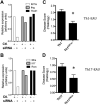
IL-27 and IL-6 signaling are important for Th1 and Th17 polarized cells, respectively, to mediate EAU. Lymph node cells from R161H mice were polarized into Th1 or Th17 phenotype. siRNAs targeting IL27Rα and IL6Rα were used to block the corresponding genes in Th1 and Th17 cells, respectively. (A) Expression of Il27ra, Ifng and Tbx21; (B) Expression of Il6ra, Il17 and Rorc by real time PCR in Th1 and Th17 polarized cells, respectively (representative of 3 independent experiments). (C,D) siRNA treated, Th1 (Th1Il27ra-) or Th17 cells (Th17Il6ra-), or cells treated with siRNA control, were transferred to WT B10.RIII recipients and disease severity was determined by histology on day 15. Data are combined from two independent experiments with total of at least 10 mice per group. *: p < 0.05.
Fig. 7
Fig. 7
IL-27p28 suppresses IL-27 and IL-6 induced Th1 and Th17 differentiation in human CD4+ T cells. (AeB) CD45RA+CD4+ naïve human T cells were isolated and polarized into Th1, with IL-12 or IL-27, and Th17, with IL-6, conditions with or without IL-27p28. Cells were harvested at day 14 for analysis. IL-27p28 significantly inhibited IL-27 induced Th1 and IL-6 induced Th17 polarization with lower production of IFN-γ and IL-17A, respectively. Data obtained from 4 independent individuals. *: p < 0.05. (C) Gene expression analysis of human IL27-induced Th1 and IL6-induced Th17 CD4+ T cells with or without IL-27p28 during polarization. (D) CD45RA+CD4+ naïve human T cells were preincubated with IL-27p28 for 2 h. Then, they were stimulated with IL-27 or IL-6 for 30 min. STAT1 and STAT3 phosphorylation was studied by intracellular staining. Representative of 3 independent individuals.

Similar articles

Cited by

References

    1. Caspi RR. A look at autoimmunity and inflammation in the eye. J Clin Invest. 2010;120:3073–83. - PMC - PubMed
    1. Ooi KG, Galatowicz G, Calder VL, Lightman SL. Cytokines and chemokines in uveitis: is there a correlation with clinical phenotype? Clin Med Res. 2006;4:294–309. - PMC - PubMed
    1. Chi W, Yang P, Li B, Wu C, Jin H, Zhu X, et al. IL-23 promotes CD4+ T cells to produce IL-17 in Vogt-Koyanagi-Harada disease. J Allergy Clin Immunol. 2007;119:1218–24. - PubMed
    1. Amadi-Obi A, Yu CR, Liu X, Mahdi RM, Clarke GL, Nussenblatt RB, et al. TH17 cells contribute to uveitis and scleritis and are expanded by IL-2 and inhibited by IL-27/STAT1. Nat Med. 2007;13:711–8. - PubMed
    1. Axtell RC, Raman C, Steinman L. Type I interferons: beneficial in Th1 and detrimental in Th17 autoimmunity. Clin Rev Allergy Immunol. 2012 - PMC - PubMed

Publication types

MeSH terms